AU-cardiac

Dilated, poorly functioning RV – PSAX view

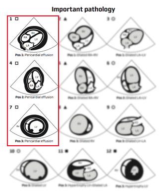
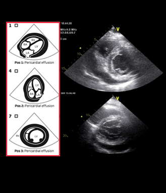
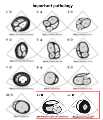
Video clips of the parasternal short axis view

Notice:

– Enlarged RV
– D-shaped left ventricle
– Paradoxial movement of the interventricular septum
– Arrows pointing to dilated right ventricle

Right ventricle enlargement and myocardial dysfunction is seen in right side myocardial infarction and pressure increase (pulmonary embolus and chronic pulmonary hypertension)

In acute enlargement the high pressure in the right ventricle will compress the left ventricle resulting in typical paradoxial interventricular septal movement and D-shape of the left ventricle

The image on the right shows hearts with enlarged and normal right ventricle – A , B and C enlarged, and D normal right ventricle.

Dilated, poorly functioning RV – PLAX view

Video clips of the parasternal long axis view

Notice
– Enlarged RV (arrows)
– RV size >2/3 of LV size except in D where there is a concomitant dilated and dysfunctioning LV

Right ventricle enlargement and myocardial dysfunction is seen in right side myocardial infarction and pressure increase (pulmonary embolus and chronic pulmonary hypertension)

Often a tricuspidal regurgitation is seen on colour Doppler and continuous wave Doppler will disclose a RV pressure increase (Advanced FATE level)

Pericardial effusion (cardiac tamponade) – characteristics

In cases with pericardial effusion different echocardiographic and clinical characteristics are observed

2D echocardiographic characteristics:

– Pericardial fluid collection
– Compression of the right-sided cavities in diastole (often not present)

Clinical characteristics:

– 5 mm wide pericardial fluid collection is within normal range
– Badly tolerated in LV hypertrophy
– Badly tolerated in postoperative cardiac surgery
– Badly tolerated in rapidly developing PE

Cardiac tamponade

Cardiac tamponade is a clinical condition where the pericardial effusion compromises the cardiac function; it may become fatal

Every physician should be able to recognise the clinical characteristics and signs of cardiac tamponade:

– Cardiac tamponade is a CLINICAL diagnosis
– Cardiac tamponade is NOT an echocardiographic diagnosis
– Pericardial effusion can often be seen with ultrasound, but the sonographic size is not
important
– The clinical presentation is of paramount importance

If therapeutic evacuation of the pericardial effusion (pericardiocentesis) stabilises the haemodynamics of the patient, the diagnosis “cardiac tamponade” was correct

Patients with clinically important PE often have concomitant pleural effusion. If so, draining of this often stabilises the patient immediately until pericardiocentesis can be performed

Image missing

Pericardial effusion – PLAX view

Video clips showing examples of pericardial effusion obtained in the parasternal long axis view

Notice:

– PLAX views
– Pericardial fluid collection (arrows)

Compression of the right-sided cavities in diastole is not obvious in these  clips

Pericardial effusion – PSAX view

Video clips showing examples of pericardial effusion obtained in the parasternal short axis view

Notice:

– PSAX views
– Pericardial fluid collection (white arrows)

Compression of the right-sided cavities in diastole is not obvious in these clips

Hypertrophic LV – PLAX view

Long axis view showing a hypertrophic left ventricle

Notice:

– Myocardial wall thickness is increased
– LV dimensions are often decreased
– Left atrium is often enlarged
– LV function is variable

Image C shows severe reduced LV dysfunction