AU-cardiac

Final remarks – Focused Cardiac Ultrasound Pathology

In these two lessons you have completed approximately 40% of a full basic FATE course

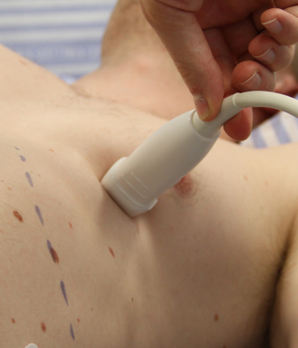
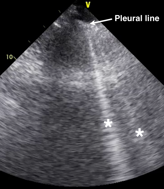
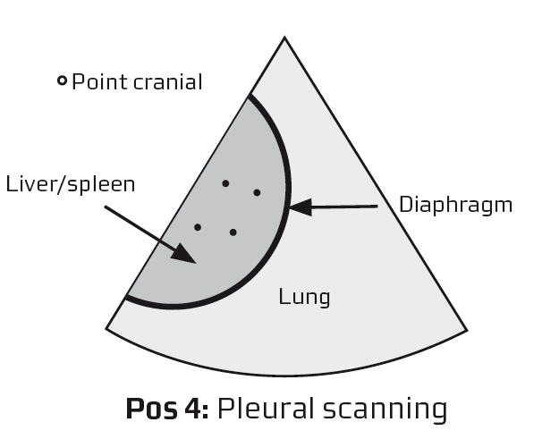
We have focused on the most important part of point-of-care cardiac ultrasound, which is 2-dimensional imaging of the most important and easiest cardiac views. In addition you have learned how to do the very important pleura scan

The patients will appreciate to be more effectively examined and treated without bias and unnecessary time delay

This is the idea of the FATE protocol

You have the basic knowledge to do this although there is much more to learn on future courses: M-mode, more pathology, cardiac systolic and diastolic function, Doppler, pressure estimation and assessment of cardiac output as the most important

Image missing
The picure shows images obtained from the four positions in the FATE examination.

Cardiac arrest – examples

Patients with cardiac arrest – 2D echocardiography

The characteristics are:

– Immobile myocardium
– Fibrillation

Possibly reversible causes for the cardiac arrest, which can be recognized with ultrasound are:

– Pericardial effusion
– Hypovolemia
– Pleural effusion
– Dilated right side (pulmonary embolus signs)
– Dilated left side

Summary – Focused Cardiac Ultrasound Pathology

You have now learned about relevant pathology which can by recognised by the FATE examination

You should be able to recognise the following conditions:
• Dilated, dysfunctional left ventricle
• Hypertrophic left ventricle with diastolic dysfunction
• Pericardial effusion / cardiac tamponade
• Dilated, dysfunctional right ventricle
• Pedunculated masses; endocarditis
• Pleural effusion
• Pulmonary edema
• Pneumothorax (presence of B-lines)
• Aorta sclerosis
• Cardiac arrest
• Enlarged atrias
• IVC abnormalities

Image missing

Limitations

Always be aware that FATE is a supplement to the clinical evaluation

FATE does not substitute a full diagnostic cardiologic echocardiographic examination

Image missing
The picure shows images obtained from the four positions in the FATE examination.

Pulmonary edema – characteristics

The 2D echocardiographic characteristics of pulmonary edema

Look for:
– B-lines

B-lines are white laser-like beams that have their origin from the pleural line and continue to the far end of the image

B-line characteristics:
– One to three is normal at the basic parts of the lungs (position 4)
– B-lines are not normal at the anterior part of thorax
– With increasing amount of lung water the number and density of the B-lines will increase
– The presence of B-lines excludes pneumothorax at the scanning point (pneumothorax can be present in other parts of the thorax)

Image missing
B-line indicated with *

Pulmonary edema – example

Multiple B-lines in a patient with pulmonary edema

Notice, B-lines start from the pleural line and extends to the the far end of the screen = 30 centimeter in depth

Image missing
Multiple B-lines obtained with a cardiac transducer

Correct tracheal intubation

The presence of lung or pleural sliding during positive pressure ventilation on both sides ensures correct placement of the endotrachial tube

The evaluation can take place anywhere on the thorax wall

Lung or pleural sliding in position 4
Notice: B-lines are present in D (normal finding in position 4 even in healthy people)

Image missing
Lung – or pleural sliding in position 4

Pneumothorax

The presence of lung sliding excludes pneumothorax

The absence of lung sliding is NOT equal to the presence of pneumothorax

Notice, lung- or pleural sliding is only possible if the two pleural blades are in contact with each other

Detailed evaluation of pneumothorax should be performed with a linear or microconvex transducer

The presence of lung- or pleural sliding excludes pneumothorax at the scanning point

The presence of just one B-line excludes pneumothorax at the scanning point

Image missing
Video clip showing both lung sliding and B-lines

If just one of the findings is present, pneumothorax can be excluded

Pleural effusion

Pleural effusion – typical clinical conditions

Should be considered in particular with the following conditions:

– All respiratory compromising conditions
– All conditions with hemodynamic instabillity
– All ICU patients
– All cardiac failure patients
– Cardio-thoracic surgery patients
– Infection
– Renal failure

Image missing