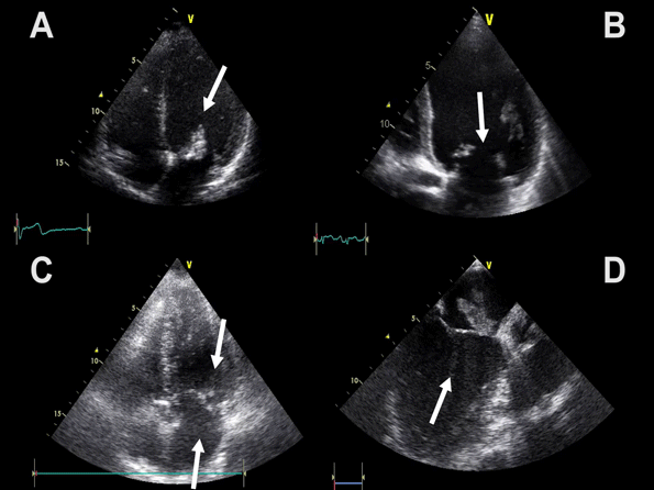
The 2D echocardiographic characteristics of pleural effusion
Look for:
– Black fluid in the thoracic cavity – above the diaphragm (fluid can appear greyish) – Atelectasis of the lung – Diaphragm unusually clearly visible – Lung sliding is absent in the phrenicocostal sinus
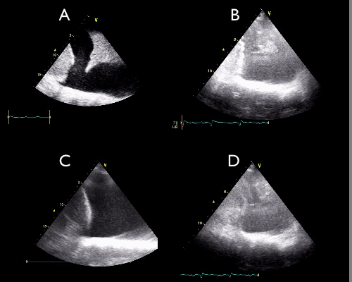
The video clips show 4 examples of pleural effusion
Notice:
– Black (or greyish) fluid collection in the thoracic cavity above the diaphragm
– Atelectasis of the lung
– Diaphragm is clearly visible
– Lung sliding is absent in the phrenicocostal sinus
Should be considered in particular with the following conditions:
– All respiratory compromising conditions – All conditions with hemodynamic instabillity – All cardiac failure patients – COPD exacercabtion – Shortness of breath, especially if arterial hypertension or hypotension is present – Volume overload, verified or suspected – Renal failure
In case of suspected pulmonary edema, ultrasound imaging of the entire thorax is indicated
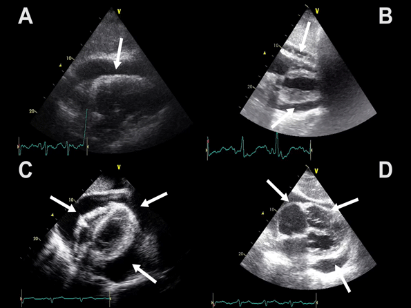
– Enlarged RV (arrows)
– RV size >2/3 of LV size
– Paradoxial movement of the interventricular septum
Right ventricle enlargement and myocardial dysfunction is seen in right side myocardial infarction and pressure increase (pulmonary embolus and chronic pulmonary hypertension)
Often a tricuspidal regurgitation is seen on colour Doppler (Advanced FATE level)
This view is not suitable for pressure measurement with continuous wave Doppler across the tricuspidal valve due to the angle of insonation (Advanced FATE level)
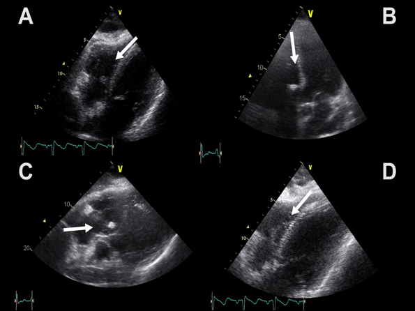
The characteristics: – Abnormal pedunculated masses – The vegetations often originate from valve leaflets but may originate from any cardiac or vascular structure – Myxomas often originate from the left atrium – Thrombus mainly originates from low flow areas, atrial appendages, areas with reduced myocardial movement, or aneurysms – Exerts their “own” movement (2D and M-mode) – Notice that pedunculated masses are not shown on the FATE card pictures
Consider in particular in the following conditions: – Endocarditis – Myxomas – Thrombus – Foreign material
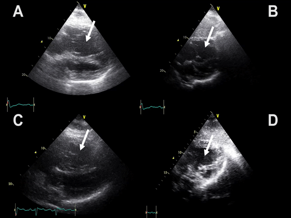
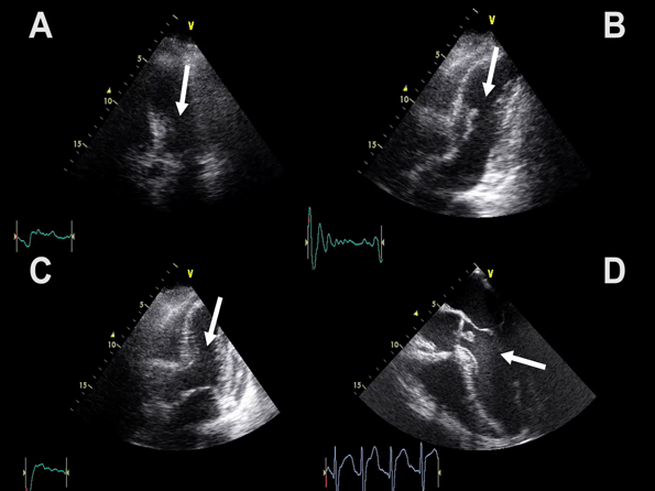
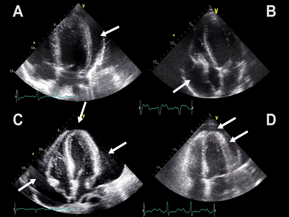
Video clips showing examples of pericardial effusion obtained in the apical 4-chamber view
Notice:
– Apical views
– Pericardial fluid collection (white arrows)
– Compression of right atrium is obvious in image B and is a
classical echocardiographic sign of tamponade, but not a prerequisite for a clinical tamponade syndrome