UGRAexp

5 – Scanning technique for the ultrasound guided greater occipital nerve block

Place the patient prone with the neck exposed.

Place a linear high-frequency probe in the axial plane across the external occipital protuberance.

Parallel shift the probe caudad to the bifid spinous process of C2. Move the probe lateral to identify the obliquus capitis inferior muscle and rotate the probe slightly to be parallel to the long axis of the muscle.

Visualize the greater occipital nerve on top of the obliquus capitis inferior muscle (see next page).

Insert the needle from the lateral end of the probe and advance the needle tip until it is in touch with the target nerve.

Inject 0.5 mL of local anaesthetic perineurally.
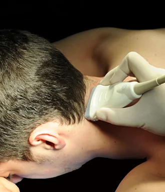
Image missing
The probe position for visualizing the bifid spinous process of vertebra C2.

7 – Sonoanatomy of the lumbar medial branch block

For the lumbar medial branch block the local anaesthetic is injected in the groove between the superior margin of the transverse process and the adjacent superior articular process.

Image missing
Local anaesthetic (yellow shadow) is injected in the groove between the transverse process (red asterisk) and the articular process (green asterisk). Spinous process (white asterisk). Needle (red).

8 – References

Finlayson RJ, Gupta G, Alhujairi M, Dugani S, Tran de QH: Cervical medial branch block: A novel technique using ultrasound guidance. Reg Anesth Pain Med 37(2): 219-23 (2012)

Shim JK, Moon JC, Yoon KB, Kim WO, Yoon DM: Ultrasound-guided lumbar medial-branch block: A clinical study with fluoroscopy control. Reg Anesth Pain Med 31: 451-54 (2006)

Image missing
Medial branches from the cervical dorsal spinal rami.

2 – Anatomy of the greater occipital nerve

The greater occipital nerve (GON) is the medial branch of the dorsal primary ramus of the second cervical nerve.

The GON innervates the muscles of the posterior neck: The semispinalis capitis and the multifidus cervicis. It also supplies cutaneous branches to the posterior scalp and meningeal branches to the posterior cranial fossa. And it supplies pain and proprioceptive branches for the first cervical nerve for innervation of the suboccipital muscles.

Image missing
The greater occipital nerve.

3 – Anatomy: The obliquus capitis inferior muscle

The greater occipital nerve curls around the lower border of the obliquus capitis inferior muscle. It is attached to the bifid spine of the axis (C2) and to the transverse process of the atlas (C1).

Image missing
The greater occipital nerve curls around the lower border of the obliquus capitis inferior muscle.

2 – Anatomy of the cervical medial branches of the dorsal spinal rami

The cervical medial branches from the dorsal spinal rami follow the deepest concavity of the articular pillars of the cervical vertebrae.

Image missing
The medial branches (yellow) from the dorsal spinal rami run posteriorly following the concavity of the middle of the articular pillar. The facet joints (green) are innervated by the medial branches. Needle (red).

3 – Anatomy of the medial branches of the lumbar dorsal spinal rami

The medial branches of the lumbar dorsal spinal rami cross the cranial margin of the transverse processes and the groove where the cranial margin of the transverse process is adjacent to the superior articular process.

Image missing
Medial branches (magenta) of the lumbar dorsal spinal rami. Needle (red). Facet joints (green).

4 – Scanning technique for the cervical medial branch block

Place the patient sitting.

Place the linear high-frequency probe in the axial plane and identify the transverse proces of vertebra C7 – the only transverse process with no anterior tubercle

Make a parallel shift cranially while counting the transverse processes until the desired level is reached

Identify the deepest concavity of the articular pillar as the most flat and echogenic part of the articular pillar (see next page)

Check for blood vessels with color Doppler

Insert the needle in-plane from the posterior end of the probe until the needle tip touches the deepest concavity of the articular pillar.

Inject 0.5 mL of local anaesthetic.

5 – Scanning technique for the lumbar medial branch block

Place the patient in the lateral decubitus position.

Place a curved array probe in the parasagittal plane and identify the superior margin of the sacrum and the transverse process of vertebra L5. Move the probe cranially while counting the transverse processes. At the target level, the probe is rotated to the axial plane and the spinous process, the articular process and the transverse process are identified.

The needle is inserted from the lateral end of the probe aiming at the groove between the superior margin of the transverse process and the superior articular proces until bony contact.

Inject 1 mL of local anaesthetic.