UGRAexp

7 – References

Børglum J, Johansen K, Christensen MD, Lenz K, Bendtsen TF, Tanggaard K, Christensen AF, Moriggl B, Jensen K: Ultrasound guided single penetration dual Injection (SPEDI) block for leg Ssrgery – a randomized controlled clinical trial. Reg Anesth Pain Med 39(1): 18-25 (2013)

Karmakar MK, Kwok WH, Ho AM, Tsang K, Chui PT, Gin T: Ultrasound-guided sciatic nerve block: Description of a new approach at the subgluteal space. Br J Anaesth 98: 390-5 (2007)

Chan VW, Nova H, Abbas S, McCartney CJ, Perlas A, Xu dQ: Ultrasound examination and localization of the sciatic nerve: A volunteer study. Anesthesiology 104: 309.14 (2006)
Image missing

2 – Anatomy of the sciatic nerve in the subgluteal space

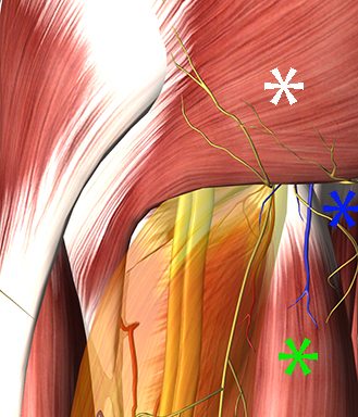
In the subgluteal space the sciatic nerve is sandwiched between the adductor magnus anteriorly and the hamstrings (long head of the biceps femoris laterally and the semitendinosus/semimembranosus medially).

Image missing
Sciatic nerve (yellow) below the transparent, yellow biceps femoris (long head), semitendinosus (green asterix), adductor magnus (blue asterix), gluteus maximus (white asterix).

3 – Scanning technique for the subgluteal approach

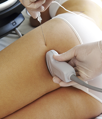
Place the patient in the lateral position

Place the low-frequency curved array probe in the gluteal crease. A linear probe can be used in lean patients

Visualize the target sciatic nerve (see next page)

Insert the needle from the lateral end of the probe with in-plane technique

Advance the needle tip to the target sciatic nerve

Inject 10-15 mL of local anaesthetic until complete perineural spread is obtained

Image missing
The ultrasound guided subgluteal sciatic nerve block.

4 – Scanning technique for the SPEDI approach

Place the patient supine.

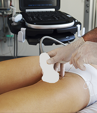
Place the low frequency curved array probe approximately 10 cm below the inguinal ligament in the femoral triangle and visualize the sciatic and saphenous nerves (see second next page).

Insert the needle from the lateral end of the probe. First advance the needle tip to the sciatic nerve and then direct it to the saphenous nerve subsartorially just lateral to the femoral artery (see second next page)

Image missing
The SPEDI block.

5 – Sonoanatomy: Subgluteal view with a high-frequency linear probe

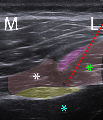
The needle is advanced from lateral to medial while avoiding piercing the posterior femoral cutaneous nerve.

Image missing
Sciatic nerve (yellow), posterior femoral cutaneous nerve (purple), long head of the biceps femoris (green asterix), semitendinosus (white asterix), adductor magnus (cyan asterix), needle (red).

6 – Sonoanatomy: SPEDI

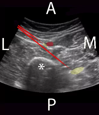
First the needle is advanced to the sciatic nerve between the gluteus maximus and adductor magnus. Then the needle is redirected to the saphenous nerve lateral to the femoral artery below the sartorius muscle in the femoral triangle.

Image missing
Sciatic nerve (large yellow shadow), saphenous nerve (small yellow shadow), femoral artery (red profile) in the femoral triangle below the sartorius muscle, femur (white asterix), needles (red).

17 – The sonoanatomy of the superficial pelvic floor

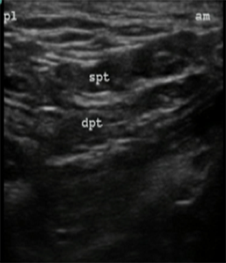
Move the probe with a parallel shift medial to the ischiocavernosus muscle in the anterior perineal triangle until the superficial and deep transverse muscles are visualized.

Image missing
Superficial transverse perineal muscle (spt), deep transverse perineal muscle (dpt), posterolateral (pl), anteromedial (am).

18 – Visualisation of the internal pudendal artery

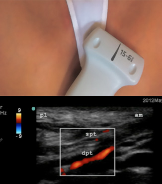
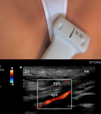
The internal pudendal artery can be visualized with Color Doppler in longitudinal view deep to the deep transverse perineal muscle. The artery can be tracked posteriorly towards the ischial tuberosity. The pudendal nerve runs alongside the internal pudendal artery either medial or lateral to the artery.

Image missing
Linear probe displaying the internal pudendal artery in longitudinal view, visualized with color Doppler. Superficial transverse perineal muscle (spt), deep transverse perineal muscle (dpt), postero-lateral (pl), antero-medial (am)

19 – Transverse view of the internal pudendal artery

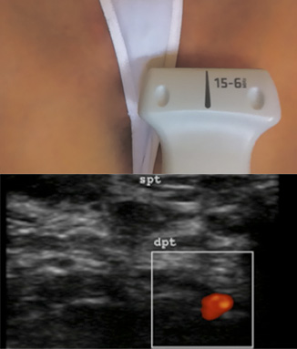
The internal pudendal artery can be visualized in transverse view with Color Doppler at the level of the ischial tuberosity.

The needle is inserted with in-plane technique from the medial end of the probe aiming just lateral to the artery. 10 mL of local anaesthetic is injected bilaterally.

Image missing
Visualization of the internal pudendal artery with Color Doppler. Superficial transverse perineal muscle (spt). Deep transverse perineal muscle (dpt).

21 – References

Parras T & Blanco R: Bloqueo pudendo ecoguiado (Ultrasound guided pudendal block). Cirugia Mayor Ambulatoria 18(1): 31-35 (2013)

http://www.asecma.org/attachments/article/162/06_18_1_FC_Parras.pdf

Parras T & Blanco R: Bloqueo perineal guiado con ultrasonidas.
Revista de anestesia regional e terapêutica da dor 70: 11-15 (2012)

http://www.anestesiaregional.com/uploads/media_items/revista-car-n-70-original.original.pdf

Image missing