UGRAexp

6 – The deep pelvic floor – the levator ani muscles

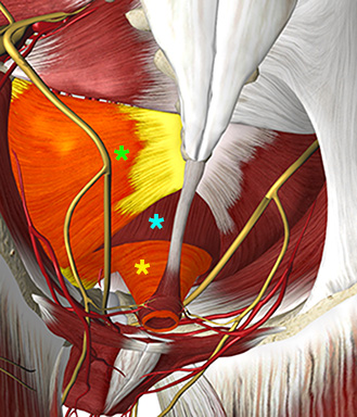
The deep pelvic floor consists of the levator ani muscles which has two major components: The pubovisceral and the iliocyccygeal muscles.

The two pubovisceral muscles are the pubococcygeus and the puborectalis muscles.

The puborectalis muscle origins from the posterior surface of the pubic bone and inserts around the rectum. It supports the contraction of the anal sphincter.

The pubocyccygeus muscle origins from the posterior surface of the superior pubic ramus and inserts on the coccyx.

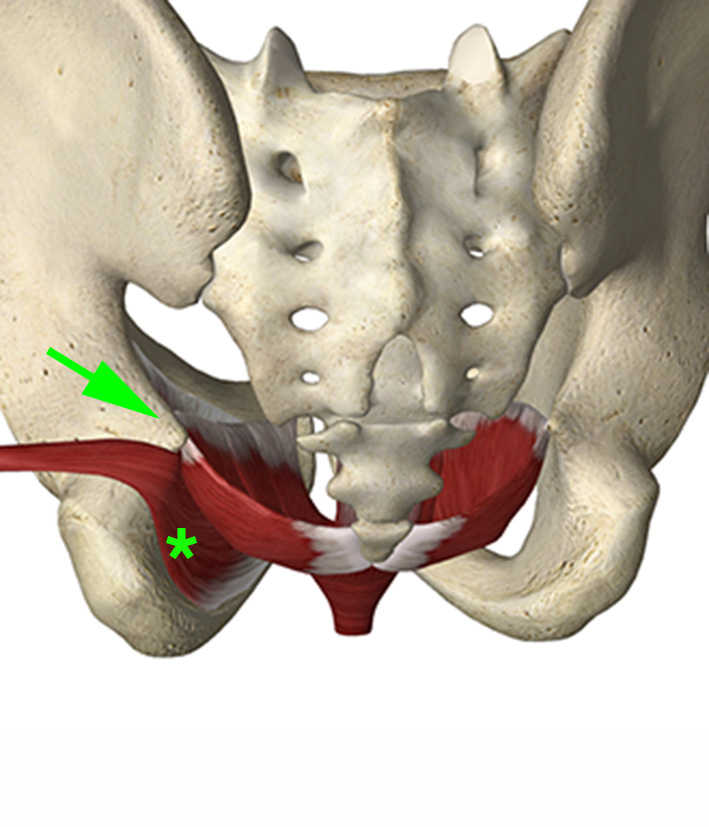
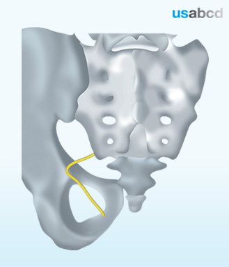
The iliococcygeal muscle origins from the ischial spine and the obturator internal muscle fascia.

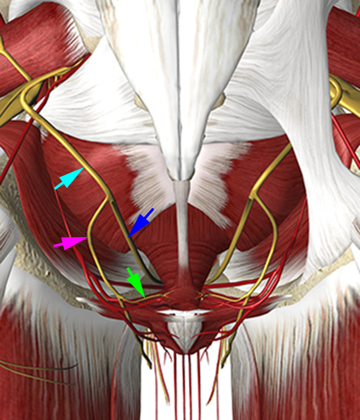
The levator ani muscles are all innervated by direct branches of the ventral primary rami of the spinal nerves S2-S4.

Image missing
The pubococcygeus muscle (cyan asterix) separates the puborectalis muscle (yellow asterix) and the iliococcygeal muscle (green asterix).

7 – The deep pelvic floor is a hammock

The three muscles of the deep pelvic floor is formed as a hammock that supports the visceral content of the pelvis.

Image missing
The levator ani (synonym: the deep pelvic floor) is formed as a hammock that origins from the ischial spine (green arrow), the fascia of the internal obturator muscle (green asterix) and the posterior aspect of the pubis. The internal obturator muscle is shown on the left side of the image.

1 – Summary

You have now completed the e-course about the expert ultrasound guided peripheral nerve blocks

You have fulfilled the following learning objectives:

– Understanding the basic anatomy and sonoanatomy of the expert nerve blocks

– Knowing how to perform each of the expert nerve blocks

– Knowing about the pitfalls

Now you are ready to join the hands-on training

Image missing

1 – Indications for the transperineal ultrasound guided pudendal nerve block

The transperineal ultrasound guided pudendal nerve block is indicated for postoperative analgesia after pelvic floor surgery including rectal extirpation.

The described technique is invented by Dr. Teresa Parras from St. Georges Hospital, London, UK, who has contributed the sonoanatomy pictures of the module.

Image missing
The pudendal nerve exits the pelvis through the greater sciatic foramen whirls around the ischial spine and enters the ischioanal fossa through the lesser sciatic foramen.

2 – Perineal area: The anterior and posterior triangles

The perineal area can be divided into two triangles: the anterior (synonym: urogenital) triangle and the posterior (synonym: anal) triangle. The base of both triangles is the superficial transverse muscles. The lateral borders of the anterior triangle are the ischiocavernosus muscles. The lateral borders of the posterior triangle is the sacrotuberous ligament.
The anterior triangle contains the root of the scrotum and penis in males and the vulva in females and it extends from the transverse perineal muscle to the pubic symphysis.
The posterior triangle contains the anal canal and its external orifice – the anus – and it extends from the transverse perineal muscle to the coccygeal bone.

Image missing
The borders of the anterior perineal triangle are the ischiocavernosus and the superficial transverse muscles – which are depicted in orange. The borders of the posterior perineal triangle are the superficial transverse muscles and the sacrotuberosus ligaments (blue color).

3 – The superficial pelvic floor

The pelvic floor can be separated into the superficial and the deep pelvic floors.

The superficial pelvic floor (synonym: the urogenital diaphragm) consists of the bulbospongiosus, ischiocavernosus, and the deep and superficial transverse perineal muscles.

The bulbospongiosus muscles (shown on the picture on page 2), the ischiocavernosus muscles and the deep and superficial transverse perineal muscles are all innervated by the deep branch of the perineal nerve from the pudendal nerve.

Image missing
The bulbospongiosus muscles have been removed exposing the deep transverse perineal muscles. The ischiocavernosus and superficial transverse perineal muscles are depicted orange on the left side of the image and the deep transverse perineal muscle is orange on the right side of the image.

4 – The innervation of the superficial pelvic floor

The superficial pelvic floor is innervated by the three pudendal nerve branches: The dorsal nerve of the penis or clitoris, the perineal nerve (synonym: the posterior scrotal nerve (or posterior labial nerve), and the inferior rectal nerve (synonym: inferior haemorhoidal nerve).

The dorsal nerve of the penis (or clitoris) is the terminal and the deepest branch of the pudendal nerve. It innervates the skin of the shaft of the penis in males and the clitoris in females.

The perineal nerve is the other terminal branch of the pudendal nerve. In males it innervates the posterior part of the skin of the scrotum. In females it innervates the posterior part of the labia and the vaginal vestibule.

The inferior rectal nerve is a branch from the perineal nerve (or directly from the pudendal nerve). It innervates the anal canal, the external anal sphincter muscle, the muscles of the superficial pelvic floor, and the puborectalis part of the levator ani muscle.

Image missing
The pudendal nerve (cyan arrow) innervates the superficial pelvic floor via its three branches: the dorsal nerve of the penis or clitoris (blue arrow), the perineal nerve (magenta arrow), and the inferior rectal nerve (green arrow).

5 – Scanning technique for the ultrasound guided greater occipital nerve block

Place the patient prone with the neck exposed.

Place a linear high-frequency probe in the axial plane across the external occipital protuberance.

Parallel shift the probe caudad to the bifid spinous process of C2. Move the probe lateral to identify the obliquus capitis inferior muscle and rotate the probe slightly to be parallel to the long axis of the muscle.

Visualize the greater occipital nerve on top of the obliquus capitis inferior muscle (see next page).

Insert the needle from the lateral end of the probe and advance the needle tip until it is in touch with the target nerve.

Inject 0.5 mL of local anaesthetic perineurally.

Image missing
The probe position for visualizing the bifid spinous process of vertebra C2.

6 – Sonoanatomy of the greater occipital nerve

The greater occipital nerve is visualized on top of the obliquus capitis inferior muscle.

The vertebral artery should be identified with color Doppler.

Image missing
Greater occipital nerve (yellow shadow), obliquus capitis muscle (white asterisk), lamina of vertebra C2 (red asterisk).

7 – References

Greher M, Moriggl B, Curatolo M, Kirchmair L, Eichenberger U: Sonographic visualization and ultrasound-guided blockade of the greater occipital nerve: A comparison of two selective techniques confirmed by anatomical dissection.
British J Anaesth 104(5): 637-42 (2010)

Image missing
The greater occiptal nerve.