DVT

Acquisition – Proximal Leg 4


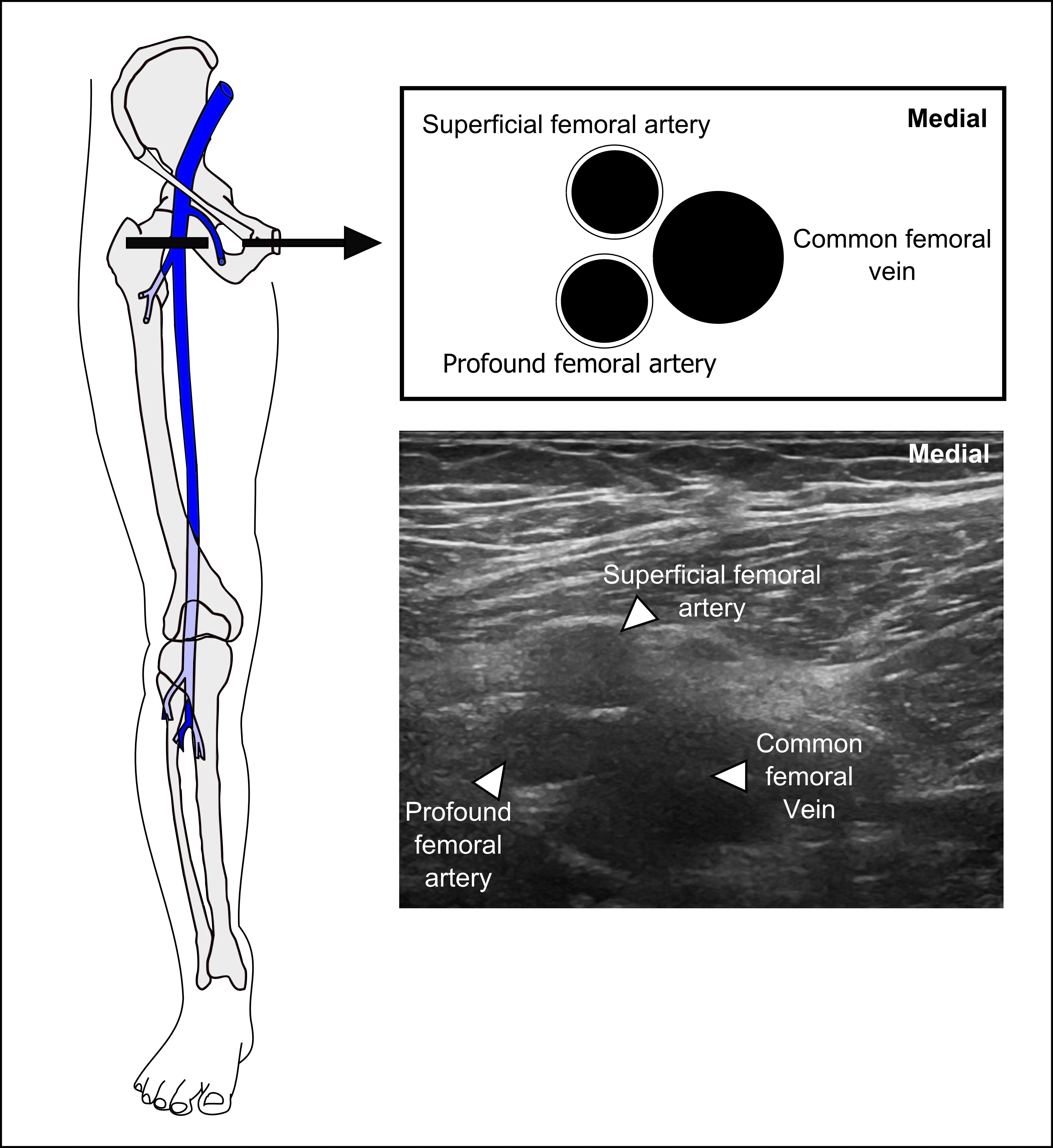
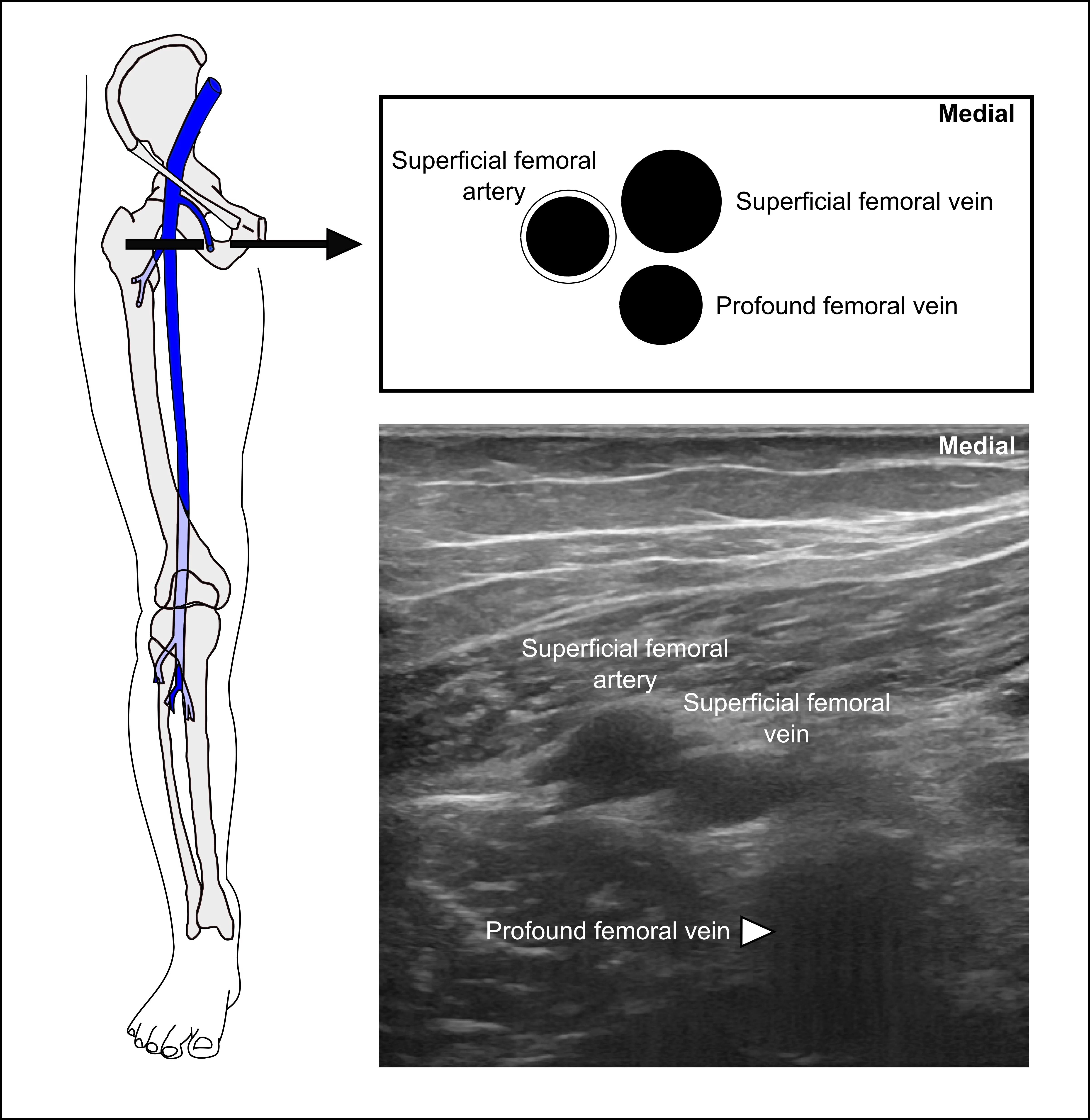
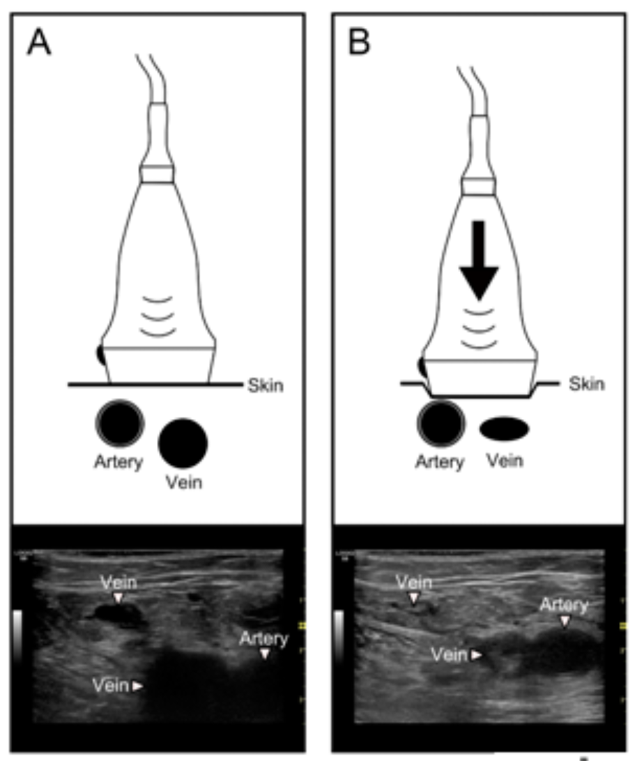
Next, the physician will sweep the ultrasound probe distally to image the bifurcation of both the common femoral vein and common femoral artery. There is anatomical variation as to which bifurcates first. The pictures here demonstrate that variability.

Note: Deep femoral artery and profound femoral artery are synonyms. Deep femoral vein and profound femoral vein are synonyms.

Image missing
Image missing

Acquisition – Proximal Leg 2


Note how the physician compresses the common femoral vein and the greater saphenous vein. The physician does not stop the compression until the lumen of both vessels have been completely compressed.

Incomplete compression is a common pitfall. Make sure you always demonstrate complete obliteration of the lumen of the veins.

Image missing
Image missing

Acquisition – Proximal Leg 1


The beginning of the POCUS evaluation for DVT occurs at the confluence of the common femoral vein and the greater saphenous vein.

  • Place the probe in the transverse orientation, just distal to the inguinal ligament.
  • The common femoral artery, common femoral vein (CFV), and the greater saphenous vein (GSV) will be seen.
  • The GSV is not considered a deep vein. However, lack of compression of the GSV at this site should be treated as a DVT.
Image missing

Proximal – Femoral Vein and Distal – Popliteal Vein


Placement of orientation marker

As with other types of ultrasound examinations, correct placement of the orientation marker on the transducer is essential in order to ensure that the orientation of the ultrasound image on the screen corresponds to the orientation of the structure scanned.

When scanning the common femoral vein and the transducer is placed on the anterior surface of the leg, the orientation marker should always be placed so it faces the patient’s right side.

When scanning the popliteal vein on the posterior side of the knee joint, the orientation marker should still be facing the patient’s right side.

Image missing
The image demonstrates how the orientation marker (OM) should be placed when scanning the patient’s right leg. A: When scanning the femoral veins, the OM is placed towards the patient’s right side. B: When scanning the popliteal veins from the posterior side of the knee, the OM is also placed towards the patient’s right side.

Acquisition


Compressibility of the deep veins will confirm the presence or absence of DVT.

Transducer: a high frequency linear transducer is usually most appropriate. A high frequency linear transducer ensures a high resolution image at the expense of penetration in terms of reduced depth

We recommend a 12-18 MHz linear probe

In obese patients, sometimes a curvilinear transducer will be needed to appropriately visualize the deep veins.

Image missing
Different ultrasound transducers or probes – the linear transducer is highlighted

Acquisition – Patient Positioning


The supine position is the ideal position for imaging the proximal leg including the common femoral and femoral veins. The leg is usually slightly flexed at the knee and the hip.

Imaging the popliteal region has numerous options:

  • Have the patient dangle the leg off the bed
  • Have the patient seated, ensuring space behind the knee
  • Have the patient supine with leg externally rotated, but flex the leg further to image behind the knee
  • Have the patient supine with their knee bent and foot flat on the bed

Indication


The following signs/symptoms are indications for evaluation for a DVT:

  • Suspicion of a lower extremity DVT
  • Leg pain
  • Leg swelling
  • Symptoms concerning for pulmonary embolism

The I-AIM Framework


The four basic steps of point-of-care DVT evaluation can be described with an I-AIM framework

I = Indication
A = Acquisition
I = Interpretation
M = Medical decision-making

Reference:

Bahner DP, Hughes D, Royall NA. I-AIM: a novel model for teaching and performing focused sonography. J Ultrasound Med. 2012 Feb; 31(2): 295-300.

Image missing

Diagnostic Accuracy of POCUS evaluation of DVT


A meta-analysis of studies evaluating the accuracy of emergency physician-performed ultrasound compared to radiology department or vascular laboratory or angiography in the diagnosis of DVT included 16 studies with 2379 patients.

Pooled test performance revealed the following test characteristics for POCUS studies for DVT:

  • Sensitivity 96.1% (95% CI 90.6-98.5%)
  • Specificity 96.8% (95% CI 94.6-98.1%)

Reference:

Pomero F, Dentali F, Borretta V, Bonzini M, Melchio R, Douketis JD, Fenoglio LM. Accuracy of emergency physician-performed ultrasonography in the diagnosis of deep-vein thrombosis: a systematic review and meta-analysis. Thromb Haemost. 2013 Jan; 109(1):137-45.

Image missing

Point-of-Care Ultrasound Protocols


2-region or 3-region Protocol

  • Limited protocol that uses compression of the femoral and popliteal regions
  • May be called “2-point” ultrasound, but this term is misleading because the proper technique is two regions rather than 2 compressions
  • No significant difference in detection of DVT in 2-region vs. 3-region protocols
  • Will miss isolated femoral vein thrombosis, present in up to 7% of patients with DVT

Extended Compression Ultrasound

  • Uses compression ultrasound from the inguinal ligament through the popliteal vein to the confluence of the calf veins
  • Will identify isolated femoral vein DVT
  • We recommend extended compression ultrasound for point-of-care ultrasound exams to improve the sensitivity of identifying isolated DVTs

Reference:

Zuker-Herman R, Ayalon Dangur I, Berant R, Sitt EC, Baskin L, Shaya Y, Shiber S. Comparison between two-point and three-point compression ultrasound for the diagnosis of deep vein thrombosis. J Thromb Thrombolysis. 2018 Jan; 45(1): 99-105.