Bates SM, et al. Diagnosis of DVT: Antithrombotic Therapy and Prevention of Thrombosis, 9th ed: American College of Chest Physicians Evidence-Based Clinical Practice Guidelines. Chest. 2012 Feb;141(2 Suppl):e351S-e418S.
Contrast Venography remains the gold standard for the diagnosis of DVT, with sensitivity and specificity of nearly 100%. It can detect DVT in the calf, iliac vessels and inferior vena. It is rarely used in the clinical environment due to its invasive nature.
D-Dimer Assay tests have sensitivity that ranges from 85-98%, depending on the pre-test probability and the type of D-Dimer assay used
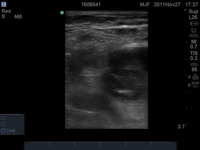
Duplex ultrasonography has a sensitivity of 93-100% and specificity of 97-100%
POCUS studies of DVT have sensitivity of 86-100% and specificity of 93-99% (1,2)
References:
1) Crisp JG, Lovato LM, Jang TB. Compression ultrasonography of the lower extremity with portable vascular ultrasonography can accurately detect deep venous thrombosis in the emergency department. Ann Emerg Med. 2010 Dec; 56(6): 601-10.
2) Kim DJ, Byyny RL, Rice CA, Faragher JP, Nordenholz KE, Haukoos JS, Liao MM, Kendall JL. Test Characteristics of Emergency Physician-Performed Limited Compression Ultrasound for Lower-Extremity Deep Vein Thrombosis. J Emerg Med. 2016 Dec; 51(6): 684-690.
• Abdominal or pelvic surgeries for cancer
• Major trauma
• Craniotomy/spinal surgery for malignant disease
• Spinal surgery with anterior approach
• Thoracic surgeries
• Major orthopedic surgery
Reference:
Heit JA, Melton LJ, Lohse CM, et al. Incidence of venous thromboembolism in hospitalized patients versus community residents. Mayo Clin Proc 2001; 76: 1102-10.
Symptomatic perioperative venous thromboembolism is a frequent complication in patients with a history of deep vein thrombosis (1).
Incidence of DVT in perioperative population:
3.6% of patients presenting for percutaneous kyphoplasty (2)
6.4% of patients with ankle fractures (3)
3.7% of patients with lower extremity fractures, and 6.2% of post-operative patients (4)
4.4% of patients presenting for gastric cancer surgery, and 7.2% of post-operative gastric cancer patients (5)
Venous thromboembolism is a factor in 5% of patients that die within 30 days postoperatively (6)
References:
1) Liem TK, Huynh TM, Moseley SE, Minjarez RC, Landry GJ, Mitchell EL, Deloughery TG, Moneta GL. Symptomatic perioperative venous thromboembolism is a frequent complication in patients with a history of deep vein thrombosis. J Vasc Surg. 2010 Sep; 52(3): 651-7.
2) Fan, Wencan MDa; Qiao, Tianzhu MDa; You, Yongqing MDb,?; Zhang, Jun MDc; Gao, Jijian MDd,? Perioperative prevalence of deep vein thrombosis in patients with percutaneous kyphoplasty, Medicine: March 2020 – Volume 99 – Issue 10 – p e19402.
3) Luo, Z., Chen, W., Li, Y. et al. Preoperative incidence and locations of deep venous thrombosis (DVT) of lower extremity following ankle fractures. Sci Rep 10, 10266 (2020).
4) Wang H, Kandemir U, Liu P, Zhang H, Wang PF, Zhang BF, Shang K, Fu YH, Ke C, Zhuang Y, Wei X, Li Z, Zhang K. Perioperative incidence and locations of deep vein thrombosis following specific isolated lower extremity fractures. Injury. 2018 Jul; 49(7): 1353-1357.
5) Osaki T, Saito H, Fukumoto Y, Kono Y, Murakami Y, Shishido Y, Kuroda H, Matsunaga T, Sato K, Hirooka Y, Fujiwara Y. Risk and incidence of perioperative deep vein thrombosis in patients undergoing gastric cancer surgery. Surg Today. 2018 May; 48(5): 525-533.
6) The Vascular Events in Noncardiac Surgery Patients Cohort Evaluation (VISION) Study Investigators. CMAJ July 29, 2019 191 (30) E830-E837.
VTE is a leading cause of preventable hospital death in the USA.
VTE is the fifth most frequent reason for unplanned hospital readmissions after surgery, overall.
Blood clots affect as many as 900,000 Americans each year leading to approximately 100,000 premature deaths.
References:
• Society of Hospital Medicine, Maynard GA, Stein JM, US Agency for Healthcare Research and Quality. Preventing hospital-acquired venous thromboembolism: a guide for effective quality improvement. Rockville, MD: Agency for Healthcare Research and Quality, US Dept. of Health and Human Services; 2008.
• Spencer F, Lessard D, Emery C, Reed G, Goldberg R. Venous thromboembolism in the outpatient setting. Arch Intern Med. 2007; 167(14): 1471-5.