DVT

Interpretation – Proximal Leg


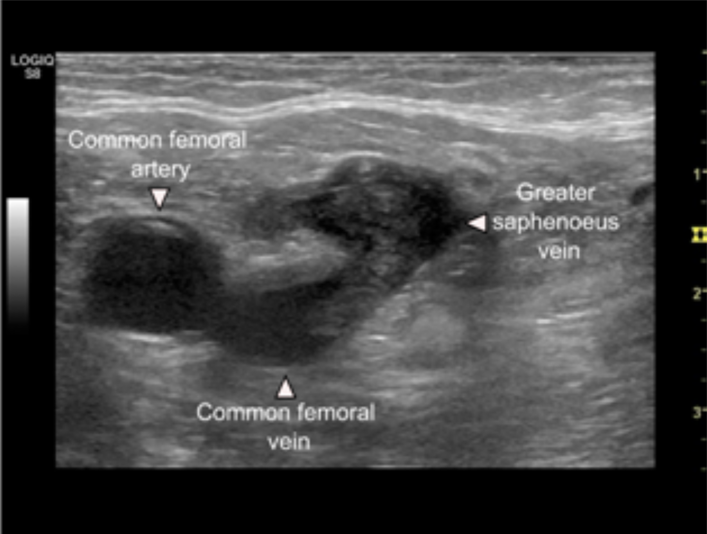
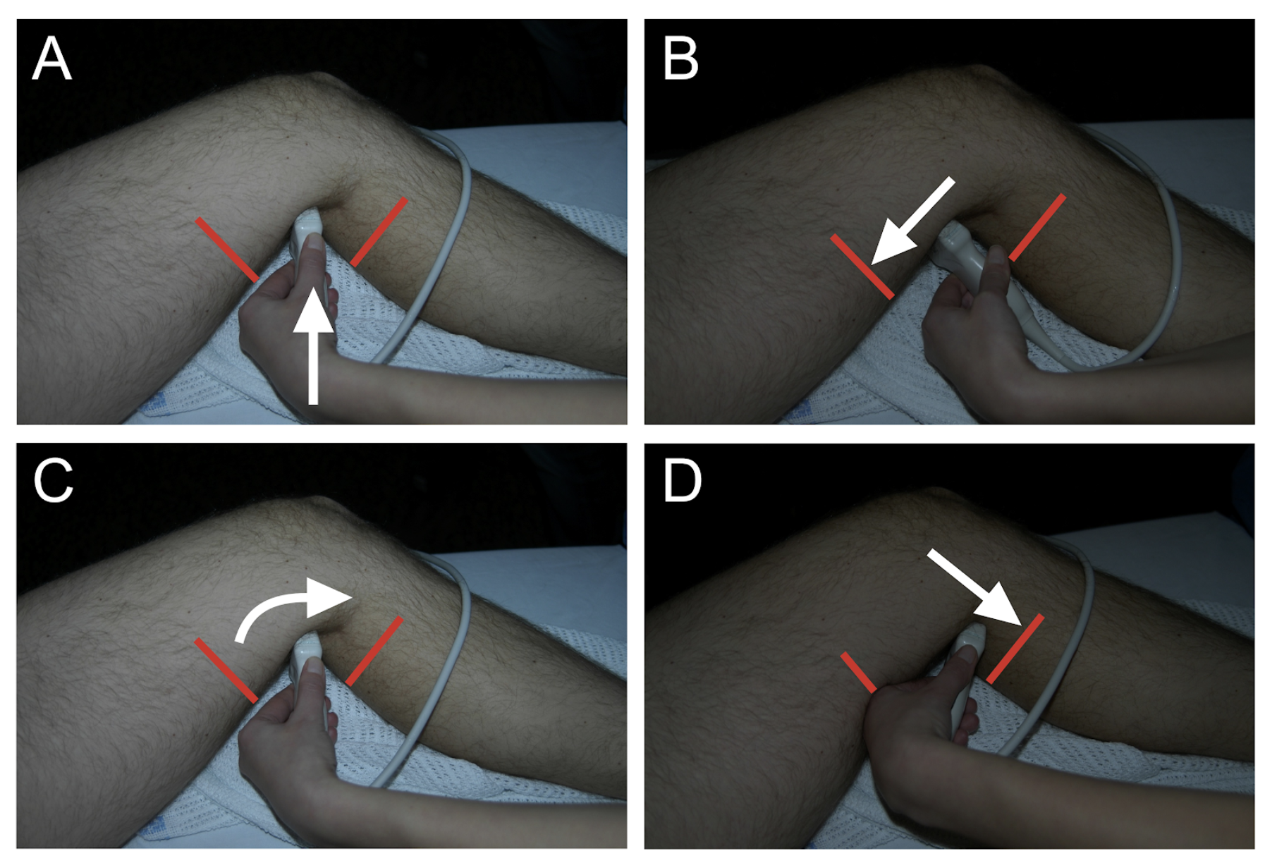
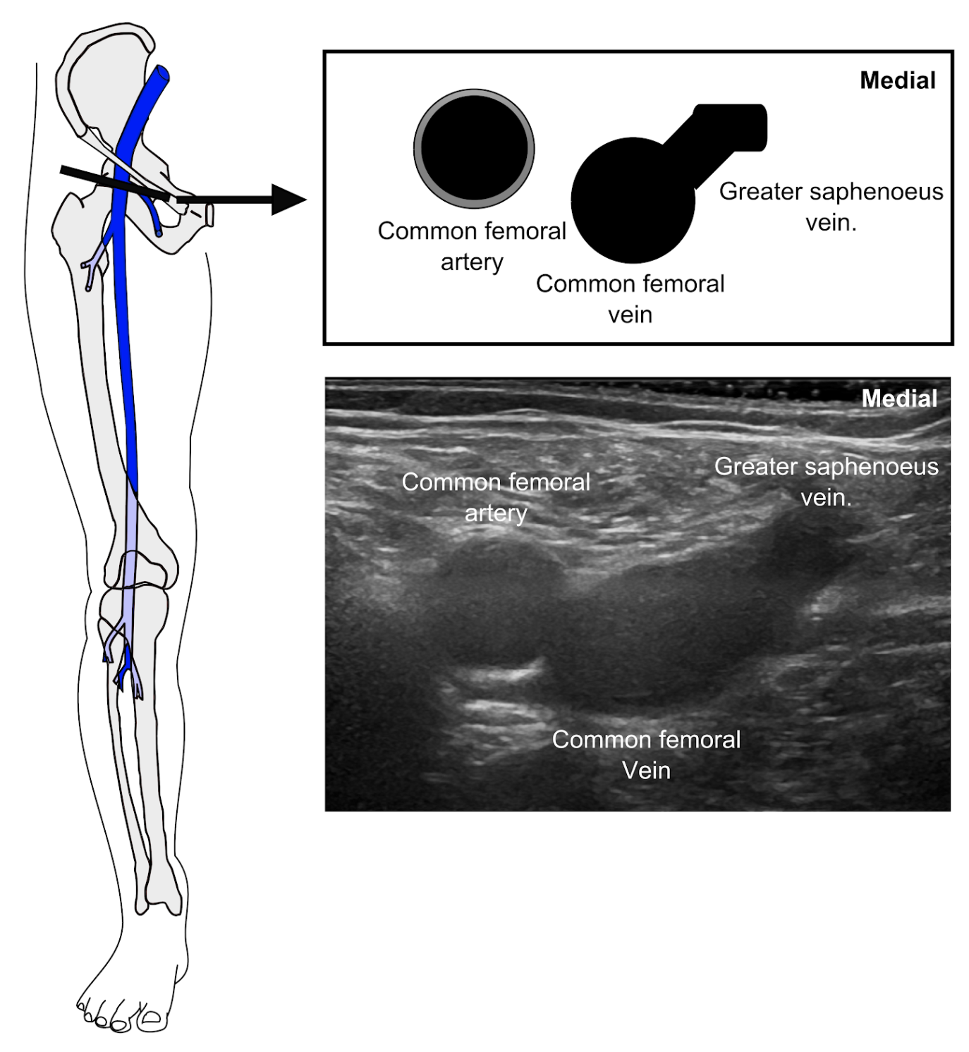
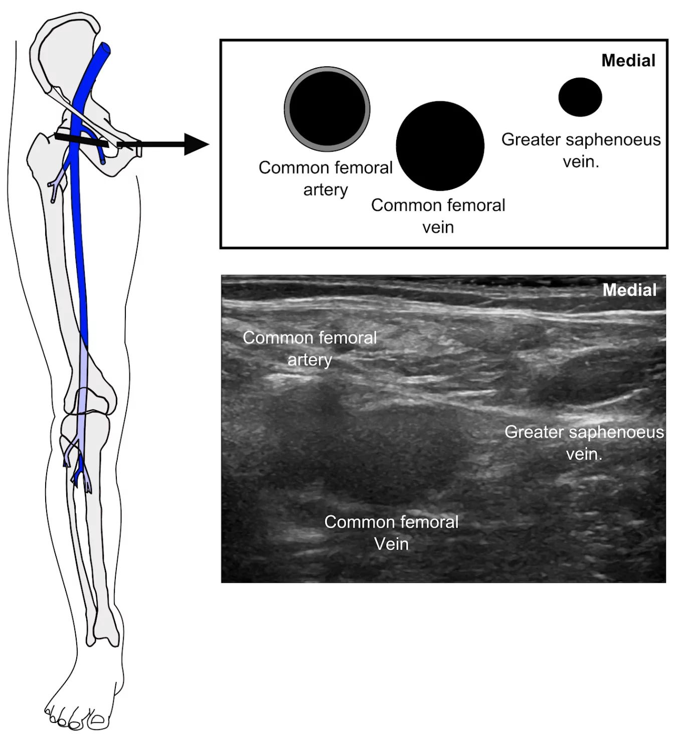
The video below is very concerning for the presence of a DVT in the common femoral vein. In this clip, the physician started compression ultrasound proximal to the intersection of the greater saphenous vein and the common femoral vein.

Image missing
Image missing
Note how the common femoral vein does not completely collapse despite adequate compression to deform the artery.

In the next video, there is heterogenous material seen at the junction of the common femoral vein and the greater saphenous vein. Even though the physician does not compress this area, the presence of heterogenous material within this region is very concerning for a DVT.
Image missing
Image missing

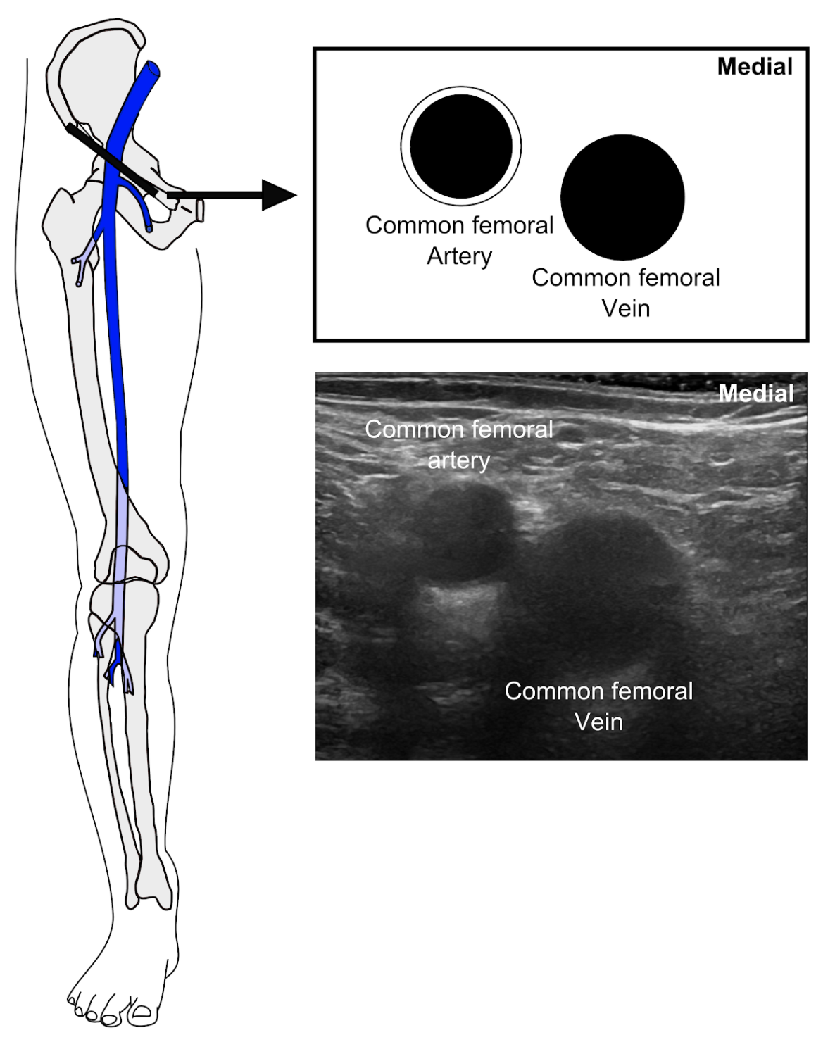
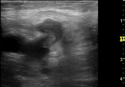
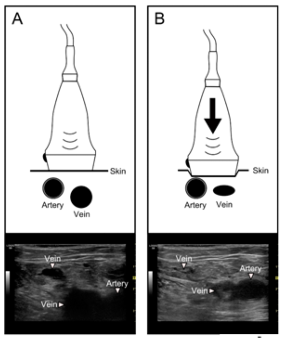
In the next video, note how the artery is somewhat compressed, but the vein does not collapse. This is highly suspicious for a DVT in the common femoral vein.

Image missing
Image missing

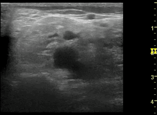
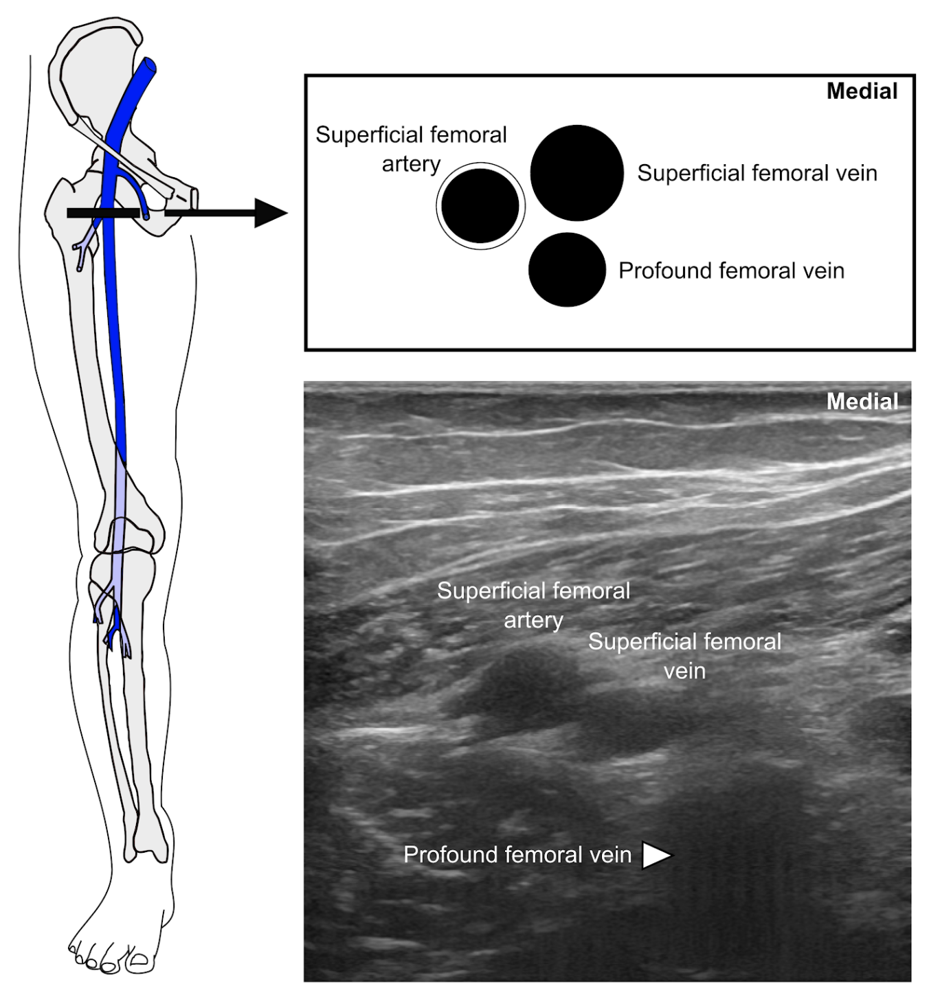
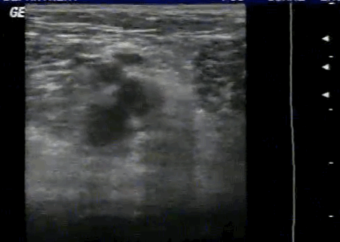
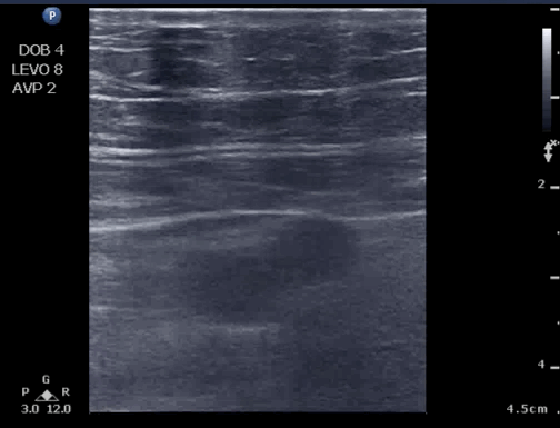
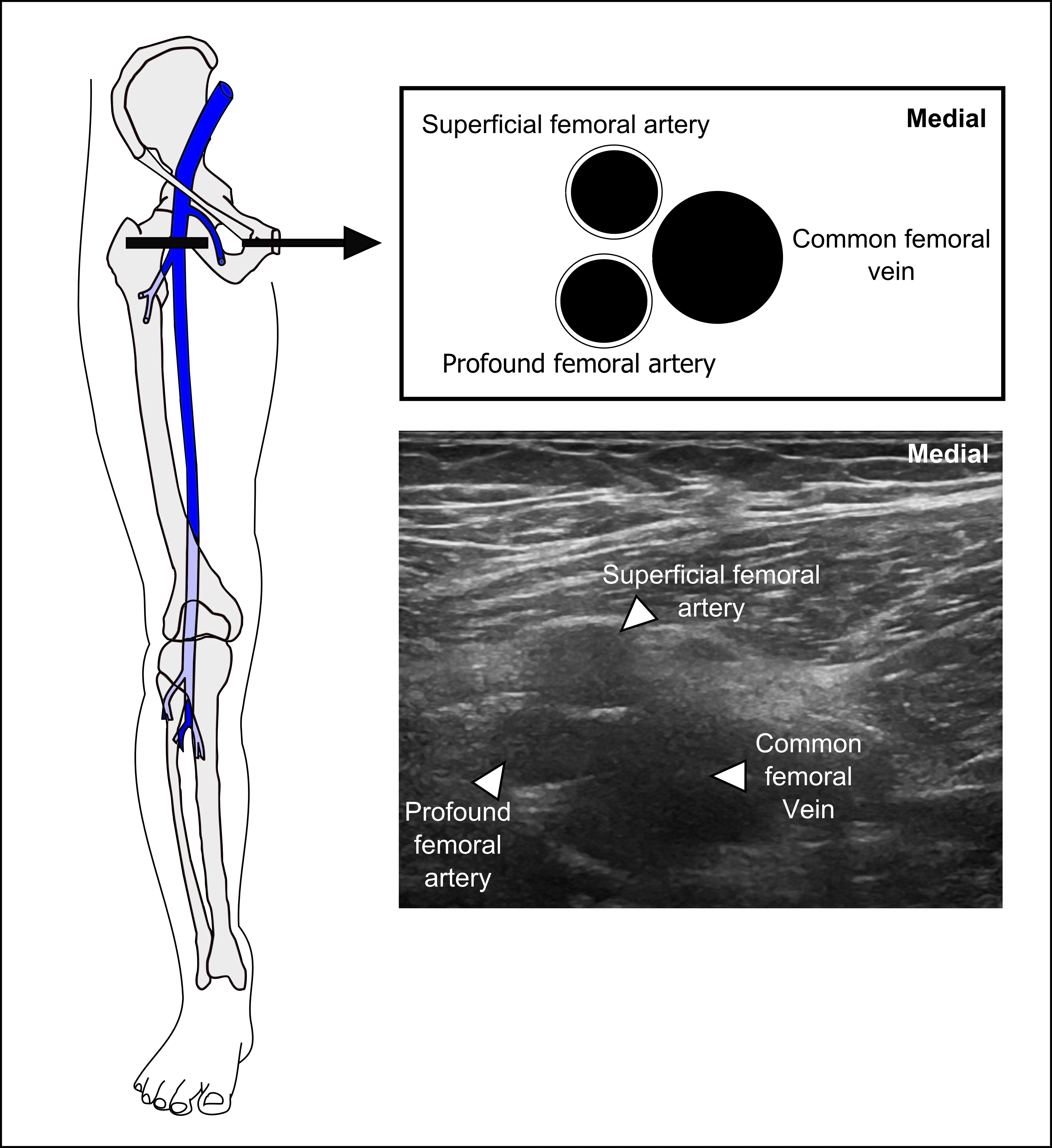
Finally, this clip demonstrates a DVT present in the femoral vein (also known as the “superficial femoral vein”). Note how the femoral artery is deformed with the pressure of compression, but the femoral vein does not collapse.

Image missing
Image missing

Interpretation


The key to interpretation is to ensure that that the physician applies adequate force on compression to cause complete coaptation of the deep vein. In cases where the vein does not completely compress, there are two main causes:

• Presence of a clot
• Inadequate pressure on the transducer

The physician can be certain that adequate pressure has been applied to the vein when the adjacent deep artery is being deformed by pressure.

Image missing

In addition to lack of compressibility of the vein, the physician may notice evidence of material in the deep veins.

Image missing
This picture demonstrates evidence of heterogeneous material in the common femoral vein and the greater saphenous vein that is consistent with a DVT.

Acquisition – Popliteal Region 3


When imaging the popliteal region, the physician will have to manipulate the probe in a small space. This will require the physician to “fan” the probe to change the probe’s angle in addition to sweeping the probe distally along the leg.

With each movement, the physician must ensure that there is complete occlusion of the deep veins with compression.

Image missing

Acquisition – Popliteal Region 4


Moving distally in the popliteal region, the physician will continue to compress the vein with each movement.

The final position will occur when the three deep calf veins are visible. It is essential that the physician compresses these veins completely to obliterate the entire lumen of the veins.

The three deep calf veins are the peroneal vein, the anterior tibial vein, and the posterior tibial vein.

Image missing

Acquisition – Popliteal Region 1


Once the proximal and mid thigh have been evaluated, move onto the popliteal region.

For this lecture, images will be shown with the patient in supine position with the knee bent and the foot on the bed. However, there are multiple positions for the patient’s leg for this position.

The initial view will start with the transducer in a location similar to a popliteal-sciatic block. The popliteal vein should be completely compressed prior to moving distally.

Image missing

Acquisition – Mid Thigh


Once adequate compression has been performed in the proximal leg, the physician continues distally down the femoral vein in the mid thigh.

At each site, the physician should perform compression of the vein to cause complete collapse of the vessel.

The physician will continue down the leg to the adductor hiatus.

Once that site is reached, the patient should change position in order for the physician to evaluate the popliteal region.

Image missing

Acquisition – Proximal Leg 5


When imaging the superficial femoral vein and the deep (profound) femoral vein, it is critical that the physician compresses both vessels. It is not sufficient to only compress one of these deep veins.
Note: despite the confusing name, the superficial femoral vein is a part of the deep venous system. Many text books and articles use the term “femoral vein” instead of “superficial femoral vein” to minimize this confusion.

Image missing

Acquisition – Proximal Leg 4


Next, the physician will sweep the ultrasound probe distally to image the bifurcation of both the common femoral vein and common femoral artery. There is anatomical variation as to which bifurcates first. The pictures here demonstrate that variability.

Note: Deep femoral artery and profound femoral artery are synonyms. Deep femoral vein and profound femoral vein are synonyms.

Image missing
Image missing