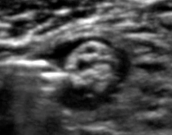
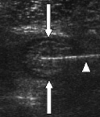
After injection of the local anaesthetic, the sciatic nerve is more easily visualised due to the contrast between the bright (hyperechoic) sciatic nerve and the background of the black (anechoic) local anaesthetic
Click on the VIDEO CLIP button to view the video
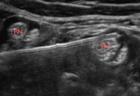
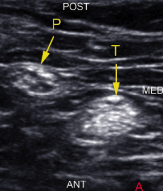
The visibility of the peroneal nerve (P) and the tibial nerve (T) is enhanced after perineural injection of a black halo
The visibility of the sciatic nerve and its branches are often reduced in the very obese patients
In obese patients it may be required to use a curved array transducer in order to perform a popliteal sciatic nerve block
However, the sciatic branches – the tibial nerve and the common peroneal nerve – are often sonographically visible near the popliteal crease, where they are usually superficially located. Even in obese patients
Click on the VIDEO CLIP button to view the video
The video shows blurred visibility of the sciatic nerve branches in an obese patient. A curved array transducer is employed to allow deeper depth of penetration of the ultrasound beam
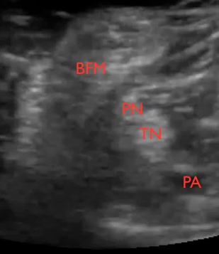
BFM = biceps femoris muscle; PN = common peroneal nerve; TN = tibial nerve; PA = popliteal artery
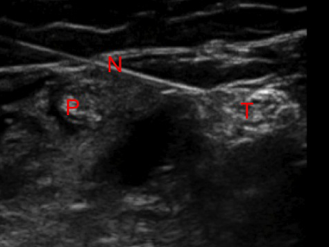
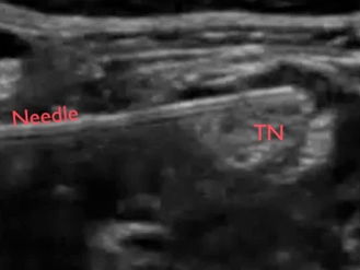
The in-plane approach to block the two branches of the sciatic nerve – the tibial nerve and the peroneal nerve – allows real-time visualisation of the needle tip, the target nerves and the perineural spread of the local anesthetic
It also gives the opportunity to relocate the needle tip, if the spread is not perfect
The real time ultrasound imaging also makes it possible to avoid piercing the peroneal nerve, when the needle is advanced towards the tibial nerve
The image simultaneously displays the target nerve (tibial nerve = T), the needle and needle tip and the peroneal nerve (P) allowing the operator to avoid piercing it when advancing the needle towards the tibial nerve
It is also possible to move the probe proximal to the bifurcation of the sciatic nerve and block the nerve guided by ultrasound at this level
Click on the VIDEO CLIP button to view the video
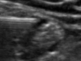
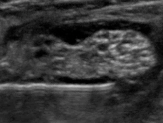
The video shows the sciatic nerve (S) in cross section just proximal to its bifurcation in the popliteal fossa. Local anesthesic (LA) is injected perineurally
PN = peroneal nerve; TN = tibial nerve
The endpoint of injection is complete perineural spread of local anaesthetic
Click on the VIDEO CLIP button to view the video
This video clip demonstrates complete perineural spread of local anaesthetic around the common peroneal nerve (P) and the tibial nerve (T) distal to the branching of the sciatic nerve in the popliteal fossa
It is easy and requires only a small volume of local anaesthetic to anaesthetise the two branches of the sciatic nerve – the tibial nerve and the common peroneal nerve – individually
The two sciatic branches are easy to access as they are more superficially located than the sciatic nerve before the bifurcation
Click on the VIDEO CLIP button to view the video
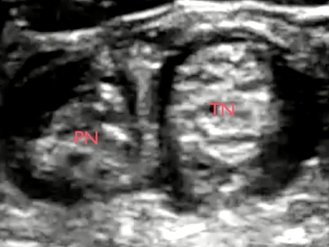
Complete perineural spread around the two sciatic nerve branches separately with only a few milliliters of local anaesthetic
Peroneal nerve (PN), tibial nerve (TN)