In the popliteal fossa the sciatic nerve splits in two branches: the tibial nerve medially and the common peroneal nerve laterally
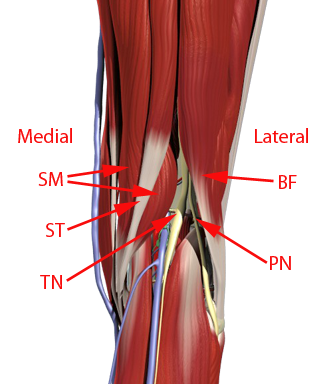
ST = semitendinosus muscle; SM = semimembranosus muscle; BF = biceps femoris muscle
In the popliteal fossa the sciatic nerve splits in two branches: the tibial nerve medially and the common peroneal nerve laterally
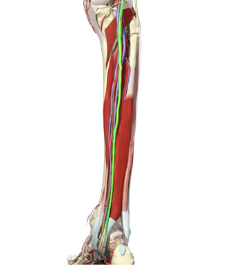
The tibial nerve runs in the posterior compartment of the leg, before it divides into the two terminal branches: the medial and the lateral plantar nerves. The two terminal branches give off cutaneous and muscular branches to the foot
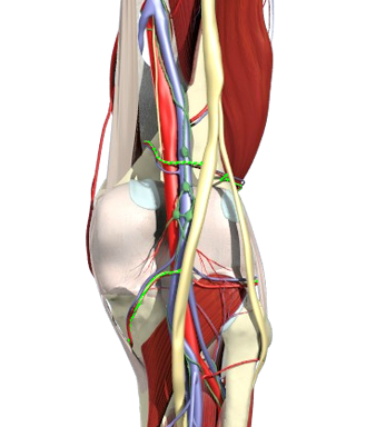
Within the popliteal fossa, the tibial nerve gives off:
– articular branches to the knee joint (the genicular nerves)
– muscular branches to the ankle flexor muscles in the posterior compartment
– a cutaneous nerve to the lateral ankle and foot – the sural nerve
In the distal part of the leg, the tibial nerve gives off:
– articular branches to the ankle joint
– a cutaneous nerve to the heel, the medial calcaneal nerve
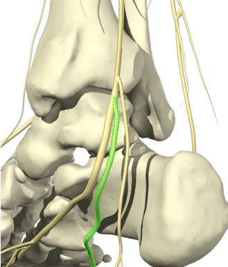
Within the popliteal fossa the tibial nerve branches off the medial genicular nerves
The lateral branches come from the peroneal nerve
The genicular nerves are sensory nerves that supply the capsule and ligaments of the knee joint
A popliteal sciatic nerve block proximal to the sciatic nerve bifurcation blocks the genicular nerves branching off the tibial nerve. I.e. the tibial innervation of the knee is blocked by a popliteal sciatic nerve block
The medial plantar nerve descends postero-inferior to the medial malleolus
It innervates the skin over the medial aspect of the sole of the foot
The nerve branches cross over to the dorsum of the toes to supply the nail beds
The medial plantar nerve distribute sensory branches to the tarsal and metatarsal joints
The medial plantar nerve supplies some of the flexor muscles of the foot
The medial plantar nerve of foot is analogous to the median nerve in the hand
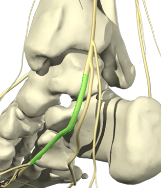
The lateral plantar nerve descends postero-inferior to the medial malleolus
It supplies some of the flexor muscles of the foot besides giving cutaneous branches to the lateral side of the sole of the foot
Branches from the lateral plantar nerve cross over to the dorsum of the toes to supply the nail bed
The lateral plantar nerve also contributes to the sensory innervation of some of the the tarsal and metatarsal joints
The lateral plantar nerve in the foot is analogous to the ulnar nerve in the hand