Lesson-Popliteal

26 – How to perform a sciatic popliteal nerve block

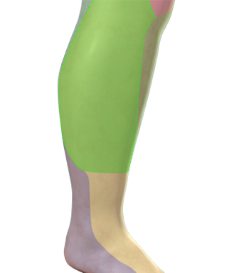
– Place the patient in the lateral position and the probe in the popliteal crease

– Visualise the tibial nerve between the popliteal artery and the probe

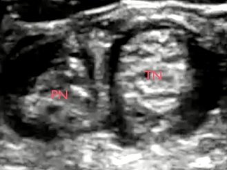
– Track the tibial nerve proximally until it unites with the peroneal nerve and the two become the sciatic nerve

– Insert the needle from the lateral end of the probe and advance the needle in-plane until the tip of the needle touches the peroneal nerve tangentially

– Inject the local anesthetic

– Relocate the tip of the needle until you obtain complete perineural spread of local anaesthetic around both the peroneal and the tibial nerve

Image missing
Top image: In-plane approach to block the two branches of the sciatic nerve.
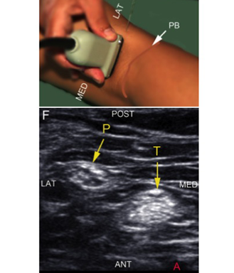
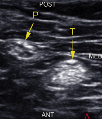
Bottom image: Sonoanatomy of the tibial and the peroneal nerve distal to the bifurcation of the sciatic nerve. (A) = popliteal artery; P = common peroneal nerve; T = tibial nerve; ant = anterior; post = posterior; lat = lateral; med = medial

27 – Use the popliteal artery as a proxy marker

– Locate the pulsating popliteal artery at the level of the popliteal crease

– Look for the tibial nerve between the artery and the skin

– Look for the common peroneal nerve lateral to the tibial nerve

– Track the tibial nerve and the common peroneal nerve proximally until they unite and become the sciatic nerve

Image missing
The image displays the peroneal nerve (P) and the tibial nerve (T) just distal to the bifurcation of the sciatic nerve in the popliteal fossa

28 – The benefit of blocking the branches after the bifurcation

It is easy and requires only a small volume of local anaesthetic to anaesthetise the two branches of the sciatic nerve – the tibial nerve and the common peroneal nerve – individually

The two sciatic branches are easy to access as they are more superficially located than the sciatic nerve before the bifurcation

Click on the VIDEO CLIP button to view the video

Image missing
Complete perineural spread around the two sciatic nerve branches separately with only a few milliliters of local anaesthetic
Peroneal nerve (PN), tibial nerve (TN)

29 – Appearance of the peroneal nerve at the bifurcation

The common peroneal nerve (P) typically presents as one single round anehoic (black) profile just before it branches off from the sciatic nerve (S)

Click on the VIDEO CLIP button to view the video

Image missing
PN common peroneal nerve; TN tibial nerve

17 – Lateral cutaneous nerve of the calf

The lateral cutaneous nerve of calf (also known as the lateral sural nerve) is a cutaneous branch of the common fibular nerve

It arises near the neck of the fibula and supplies skin over the lateral aspect of the leg just distal to the knee

Image missing
The green colour depicts the cutaneous distribution of the lateral cutaneous nerve of the calf

18 – The superficial peroneal nerve – a terminal branch of the common peroneal nerve

The superficial fibular nerve is the smallest of the two terminal branches of the common fibular nerve

It innervates the muscles of the lateral compartment of the leg

It supplies cutaneous branches to the lateral aspect of the distal half of leg

The branches of the superficial peroneal nerve supply the dorsum of the foot and the toes

Image missing
The green colour depicts the superficial peroneal nerve

19 – Cutaneous distribution of the superficial peroneal nerve

The superficial fibular nerve emerges through the fibular musculature about halfway down the leg

It innervates the dorsum of the foot and the toes – except the interspace between the first and the second toe

Image missing
The green colour depicts the cutaneous distribution of the superficial peroneal nerve

20 – The deep peroneal nerve

The deep peroneal nerve is the biggest of the two terminal branches of the common peroneal nerve

The deep peroneal nerve descends within the anterior crural compartment and supplies all the muscles of this compartment

It crosses in front of the ankle joint, giving an articular branch to the ankle joint, before the terminal branches innervate the dorsum of the foot, lateral to the dorsalis pedis artery

It innervates the skin of the interspace between the 1st and 2nd toes

It also innervates the digital extensor muscles and some of the tarsal and tarsometatarsal joints

Image missing
The green colour depicts the deep perineal nerve