Lesson-CardiacUS
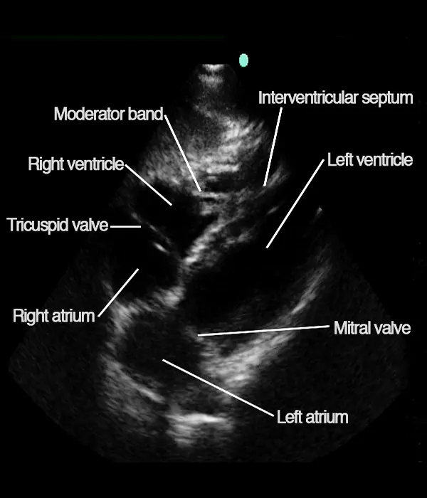
The subcostal 4-chamber view – identifying structures
The following important structures are seen in the subcostal 4-chamber view:
• Left atrium
• Left ventricle
• Mitral valve
• Right atrium
• Right ventricle
• Tricuspid valve
• Septum intraventriculare
The following focused questions can be assessed:
• Is a pericardial effusion present?
• Is the left ventricular function reduced?
• Are signs of pulmonary embolism present?
A good subcostal 4-chamber view is valuable for assessing all three of the above mentioned focused questions – but especially for pericardial effusion assessment.
The subcostal 4-chamber view – tips and tricks
Tips and tricks
In order to obtain the subcostal acoustic window, sufficient pressure needs to be applied to the transducer so the ultrasound waves can travel under the rib cage as seen in the image below.
Asking the patient to bend his legs often helps as this causes relaxation of the abdominal muscles allowing for correct transducer placement.
Asking the patient to take a deep inspiration often improves image quality because the heart is displaced towards the abdomen and the transducer (remember to tilt the tail of the transducer upwards).
Once you have found an acoustic window, try optimizing the image (tilting and rotating) so the cavities are fully open. Often, a 10 degree counter-clockwise rotation is necessary.
If you get a lung-shadow across the apex, try sliding the transducer a little towards the patient’s right side.
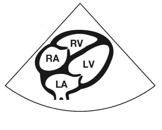
RA: Right atrium
RV: Right ventricle
LA: Left atrium
LV: Left ventricle
