You have now learned how to answer the following focused questions:
• Is a pericardial fluid collection present?
• Is the left ventricle systolic function reduced?
• Are signs of aortic stenosis present?
• Are signs of right heart strain present?
• Are signs of hypovolemia present?
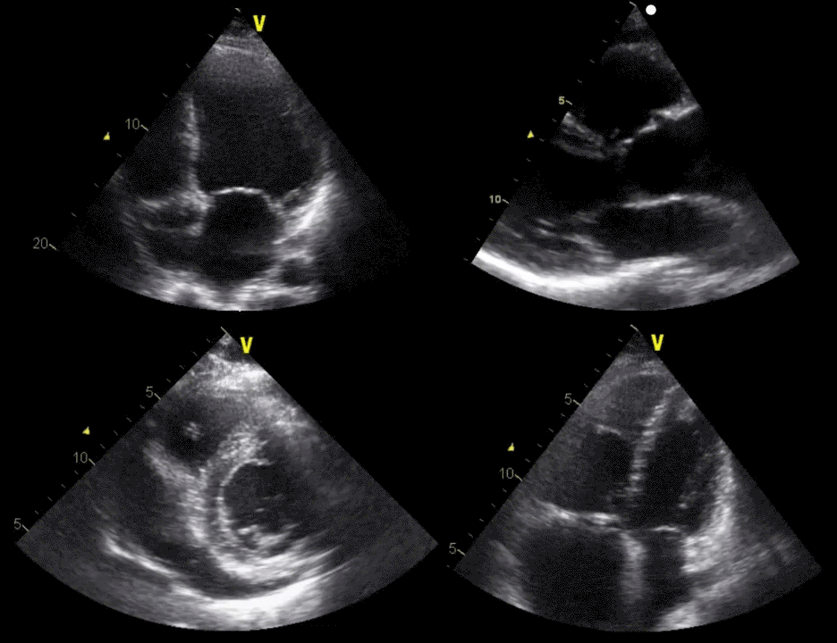
The four cardiac pathologies demonstrated. Upper left: reduced left ventricle systolic function; upper right: ascending aortic dissection; lower left: pericardial fluid collection; lower right: pulmonary embolism.
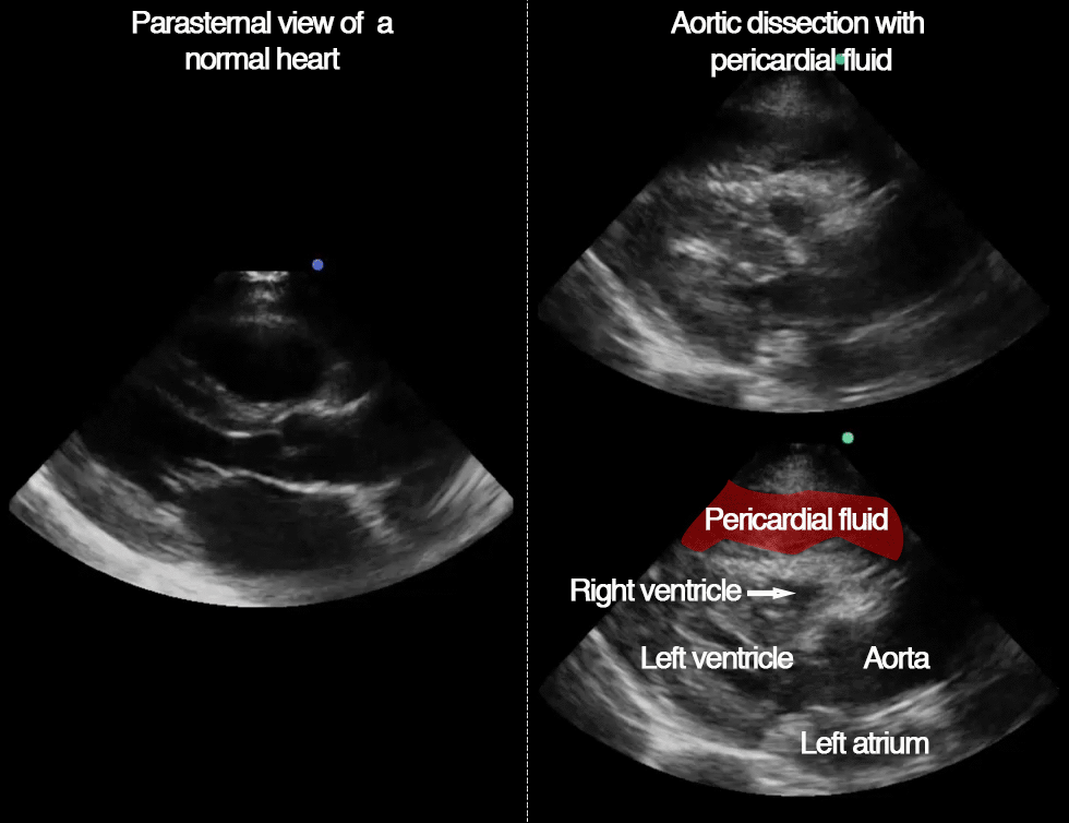
In a patient with a suspected type A aortic dissection, the presence of pericardial fluid collection is highly suggestive of dissection.
Tips
If lacking good parasternal images, any finding of pericardial fluid collection in a patient with the relevant symptoms should trigger triage to the invasive heart center.
Pericardial effusion in the PLAX view. Normal heart to the left.
Facts
• The ascending aortic diameter is highly variable and increases with increasing body-surface area and age.
• An ascending aortic maximum diameter >5 cm is always pathological – regardless of age and body-size.
• An aortic maximum diameter <3.5 cm makes aortic dissection unlikely.
• Approximate distribution of size at the time of dissection:
<4 cm: 5% , 4-5 cm: 35%, >5 cm: 60%.
Tips
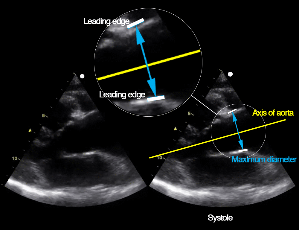
• The ascending aorta is measured at the point of largest diameter.
• Precise measurements are done perpendicular to a line through the centre of aorta during systole.
• The aortic diameter is measured from the leading edge of the anterior aortic wall to leading edge of the posterior aortic wall.
Facts
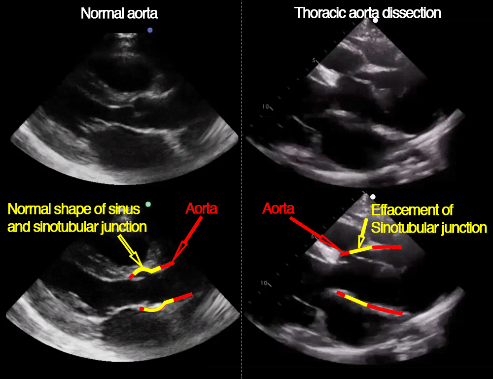
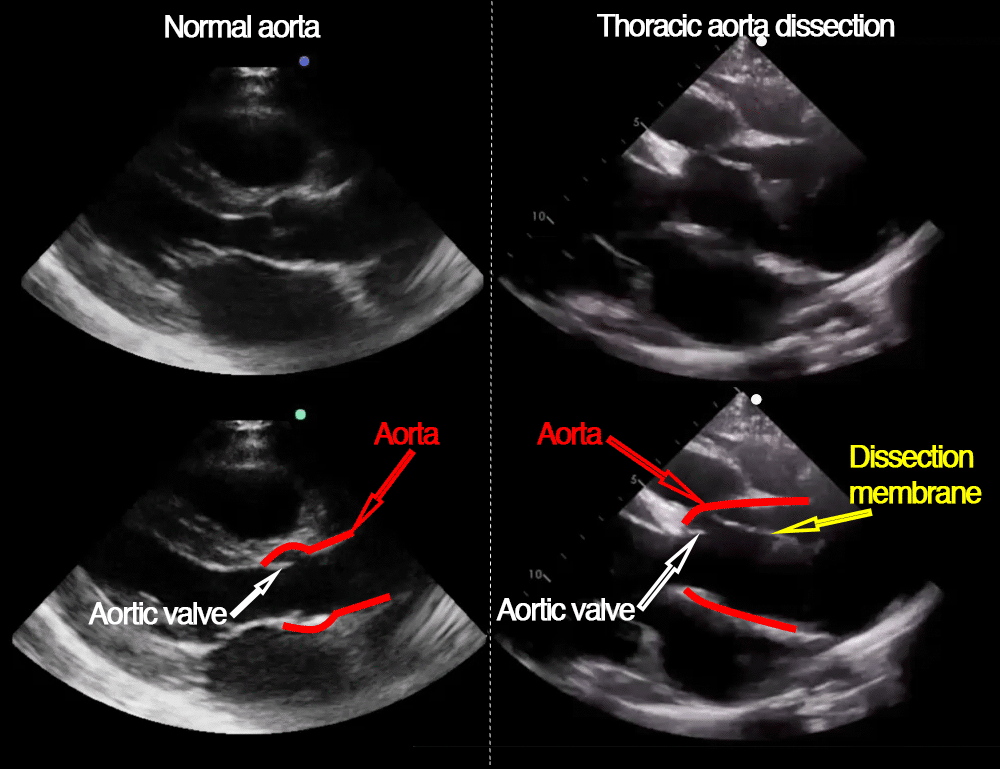
The ascending aorta can be divided into sinus valsalva, just above the aortic valve, and the more distal tubular part.
With normal anatomy, there is a narrowing of the aorta at the sino-tubular junction causing the characteristic shape of the most proximal part of the ascending aorta.
In most cases of type A aortic dissection, this characteristic shape is lost because of dilation at the sino-tubular junction.
Facts
A dissection membrane will not always be visible with transthoracic ultrasonography in case of type A aortic dissection, but when present it is highly suggestive of dissection.
Facts
No ultrasonographic finding can rule out aortic dissection.
However, in combination with relevant symptoms, four findings are highly suggestive of type A aortic dissection:
• the presence of a dissection membrane
• loss of sino-tubular junction
• largest ascending aorta diameter >5 cm
• pericardial fluid collection
Two findings reduce a suspicion of type A aortic dissection:
• preserved sino-tubular junction
• largest ascending aortic diameter <3.5 cm
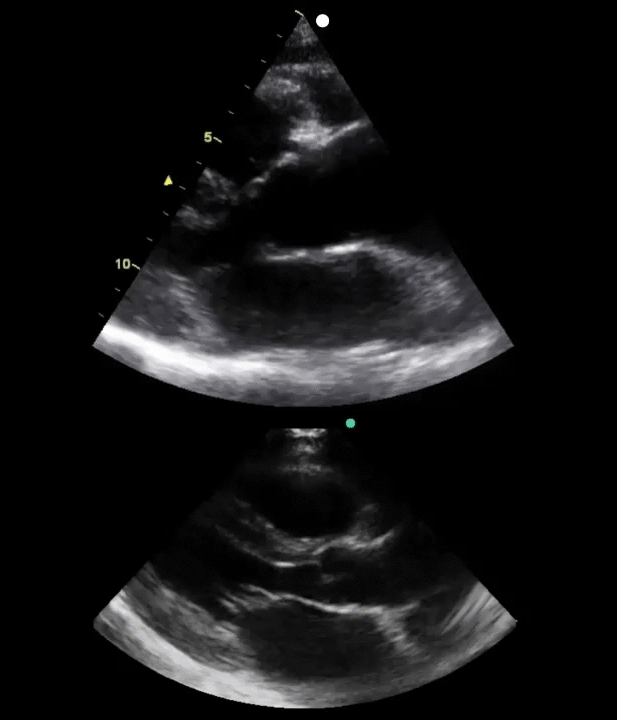
Top: Type A thoracic aortic dissection
Bottom: Parasternal view of a normal heart
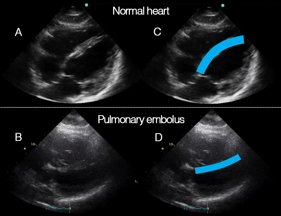
In healthy people, the septum bulges from the left ventricle into the right because of the higher pressure in the left ventricle.
When the pressure in the right ventricle increases to levels higher than in the left ventricle, it bulges from the right to the left.
Depending on the pulmonary pressure, this septum-shift may be seen only in diastole or in both systole and diastole. In the video below it is seen during diastole in the heart with pulmonary embolism.
A pulmonary embolism that causes severe circulatory failure is unlikely without this septum-shift.
Tips
Practice looking for a septum-shift towards the left ventricle.
The septum is a very hyperechogenic structure and can often be visualised even in poor images.
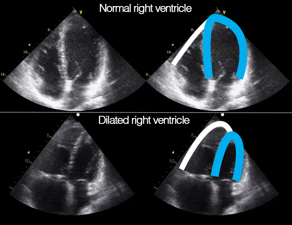
Compare the size of the right ventricle to that of the left ventricle by eyeballing.
If the right ventricle diameter is equal to or larger than the left ventricle, it is dilated.
In a patient with the relevant symptoms, this supports the diagnosis of pulmonary embolism.
Notice that with dilation and elevated right ventricular pressure, the right ventricle can become apex forming as seen in the image to the right.
Tips
In the subcostal view, the width at the base of the right ventricle can look enlarged if the transducer is not rotated correctly.
Enlargement of the right ventricle is not a specific sign of pulmonary embolism, but is also seen in chronic pulmonary hypertension and right coronary artery occlusion.
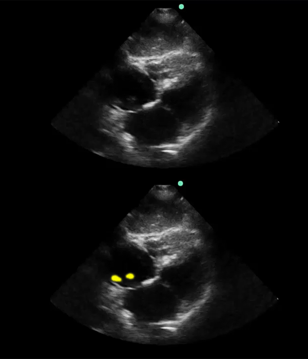
Visible intracavitary thrombolus is not a frequent sign in pulmunary embolism – but if it is present it is highly specific and associated with high risk.
Visible intracavitary thromboembolus can be seen both in the right atrium and right ventricle.
Tips
Patients with symptoms of pulmonary embolism and visible intracavitary thromboembolus should be triaged directly to invasive heart center.
Subcostal 4-chamber views with visible intracavitary thromboembolus in the right atrium (highlighted in yellow at the bottom).