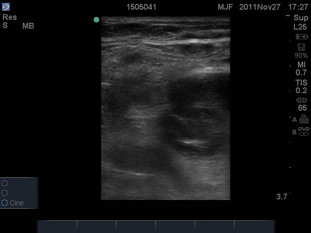
The supine position is the ideal position for imaging the proximal leg including the common femoral and femoral veins. The leg is usually slightly flexed at the knee and the hip.
Imaging the popliteal region has numerous options:
Have the patient dangle the leg off the bed
Have the patient seated, ensuring space behind the knee
Have the patient supine with leg externally rotated, but flex the leg further to image behind the knee
Have the patient supine with their knee bent and foot flat on the bed
A meta-analysis of studies evaluating the accuracy of emergency physician-performed ultrasound compared to radiology department or vascular laboratory or angiography in the diagnosis of DVT included 16 studies with 2379 patients.
Pooled test performance revealed the following test characteristics for POCUS studies for DVT:
Sensitivity 96.1% (95% CI 90.6-98.5%)
Specificity 96.8% (95% CI 94.6-98.1%)
Reference:
Pomero F, Dentali F, Borretta V, Bonzini M, Melchio R, Douketis JD, Fenoglio LM. Accuracy of emergency physician-performed ultrasonography in the diagnosis of deep-vein thrombosis: a systematic review and meta-analysis. Thromb Haemost. 2013 Jan; 109(1):137-45.
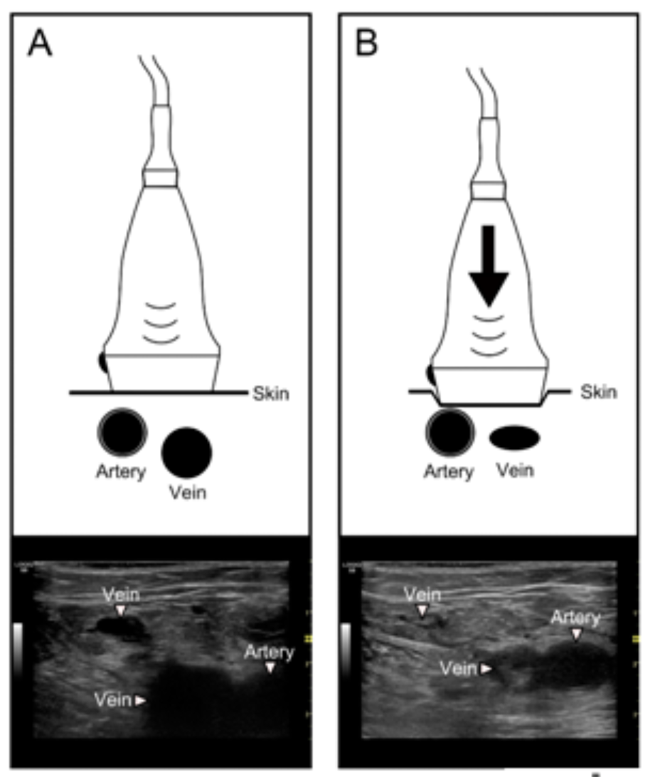
Limited protocol that uses compression of the femoral and popliteal regions
May be called “2-point” ultrasound, but this term is misleading because the proper technique is two regions rather than 2 compressions
No significant difference in detection of DVT in 2-region vs. 3-region protocols
Will miss isolated femoral vein thrombosis, present in up to 7% of patients with DVT
Extended Compression Ultrasound
Uses compression ultrasound from the inguinal ligament through the popliteal vein to the confluence of the calf veins
Will identify isolated femoral vein DVT
We recommend extended compression ultrasound for point-of-care ultrasound exams to improve the sensitivity of identifying isolated DVTs
Reference:
Zuker-Herman R, Ayalon Dangur I, Berant R, Sitt EC, Baskin L, Shaya Y, Shiber S. Comparison between two-point and three-point compression ultrasound for the diagnosis of deep vein thrombosis. J Thromb Thrombolysis. 2018 Jan; 45(1): 99-105.
Bates SM, et al. Diagnosis of DVT: Antithrombotic Therapy and Prevention of Thrombosis, 9th ed: American College of Chest Physicians Evidence-Based Clinical Practice Guidelines. Chest. 2012 Feb;141(2 Suppl):e351S-e418S.
Contrast Venography remains the gold standard for the diagnosis of DVT, with sensitivity and specificity of nearly 100%. It can detect DVT in the calf, iliac vessels and inferior vena. It is rarely used in the clinical environment due to its invasive nature.
D-Dimer Assay tests have sensitivity that ranges from 85-98%, depending on the pre-test probability and the type of D-Dimer assay used
Duplex ultrasonography has a sensitivity of 93-100% and specificity of 97-100%
POCUS studies of DVT have sensitivity of 86-100% and specificity of 93-99% (1,2)
References:
1) Crisp JG, Lovato LM, Jang TB. Compression ultrasonography of the lower extremity with portable vascular ultrasonography can accurately detect deep venous thrombosis in the emergency department. Ann Emerg Med. 2010 Dec; 56(6): 601-10.
2) Kim DJ, Byyny RL, Rice CA, Faragher JP, Nordenholz KE, Haukoos JS, Liao MM, Kendall JL. Test Characteristics of Emergency Physician-Performed Limited Compression Ultrasound for Lower-Extremity Deep Vein Thrombosis. J Emerg Med. 2016 Dec; 51(6): 684-690.