Emergency
Perisplenic view: Identifying structures Copy BUA
In the perisplenic view, the following structures are seen:
• Diaphragm
• Spleen
• Kidney
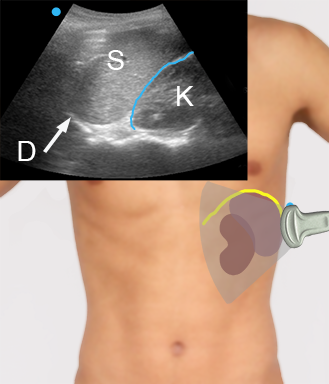
D: Diaphragm
S: Spleen
K: Kidney
The splenorenal recess is marked in blue.
Obtaining the perisplenic view Copy BUA
The perisplenic view is obtained in the same way as perihepatic view.
Be aware that a slight rotation on the transducer in order to avoid rib shadowing should be done with a clockwise rotation as opposed to the counter-clockwise rotation in the perihepatic view.
Perihepatic view: Identifying structures Copy BUA
In the perihepatic view, the following structures are seen:
• Diaphragm
• Liver
• Kidney
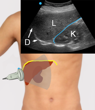
D: Diapgragm, L: Liver, K: Kidney. Morison’s pouch is marked in blue
Abdominal views when looking for free fluid Copy BUA
To look for free fluid in the abdomen, four views are relevant:
• The perihepatic view
• The perisplenic view
• The pelvic views – transverse and longitudinal views
• The peri-intestinal view(s)
The next sections will take you through how to obtain these views
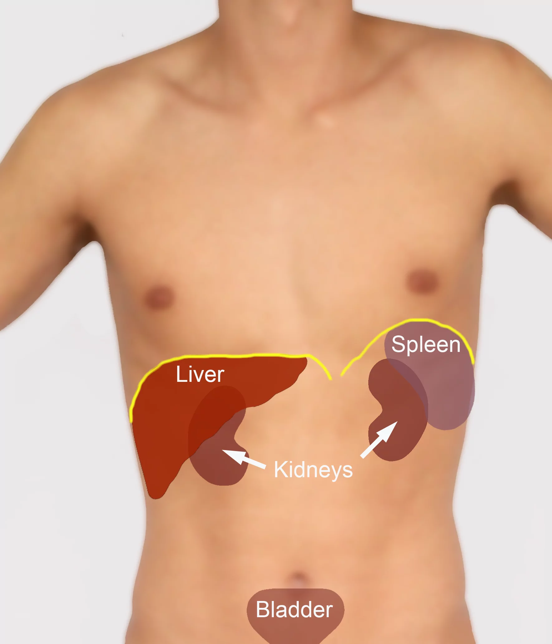
Perihepatic view: Diaphragm, liver and kidney
Perisplenic view: Diaphragm, spleen and kidney
Pelvic views: Bladder and rectum – and in women also uterus (not demonstrated)
Peri-intestinal view
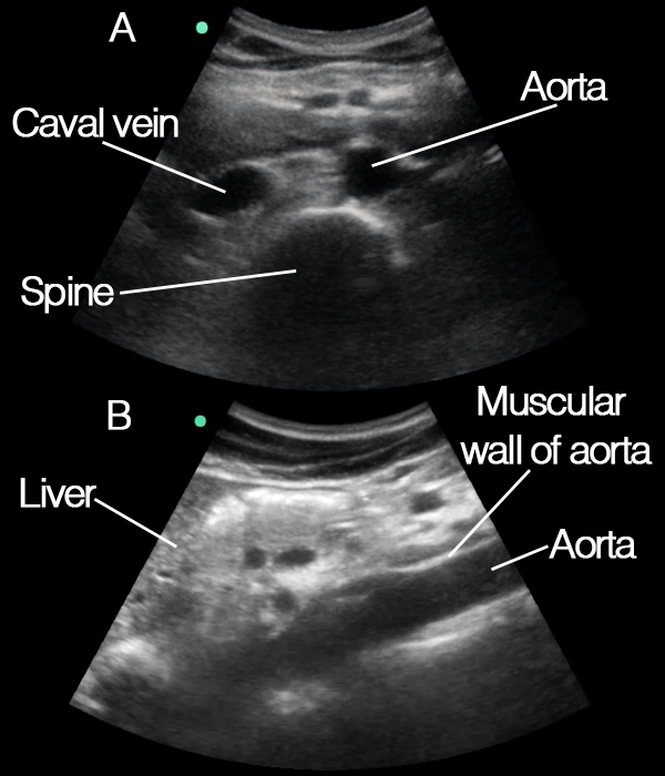
Slettes Abdominal aorta: Identifying structures Copy BUA
In the transverse aortic view, the following structures are seen:
• Aorta
• Inferior vena cava
• Columna vertebralis
In the longitudinal abdomnial aortic view, the following structures are seen:
• Aorta
• Liver (in the upper part of the abdomen)
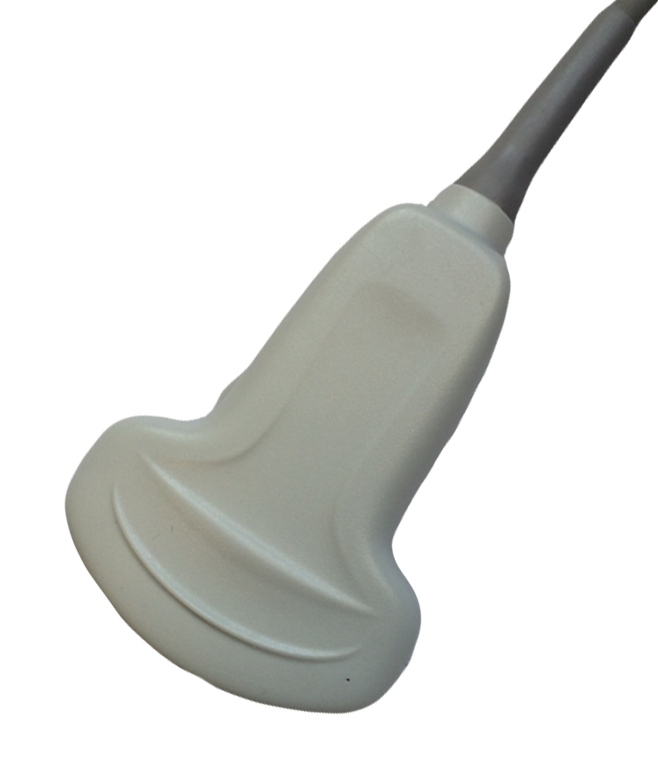
The transducer for focused ultrasonography of the abdomen Copy BUA
For focused ultrasonography of the abdomen a lower frequency curved (abdominal) transducer is used.
A lower-frequency curved transducer (abdominal) permits the deep penetration that is needed for evaluating deeper structures and has a wide footprint that allows a good overview.
If no abdominal transducer is available, a cardiac transducer can be used, but the small footprint of this transducer does not offer as good an overview.
Tips and tricks
The ‘abdominal’ transducer can be used for all lung and abdominal examinations.
Summary – focused cardiac questions
You have now learned how to answer the following focused questions:
• Is a pericardial fluid collection present?
• Is the left ventricle systolic function reduced?
• Are signs of aortic stenosis present?
• Are signs of right heart strain present?
• Are signs of hypovolemia present?