Emergency

Which transducer?


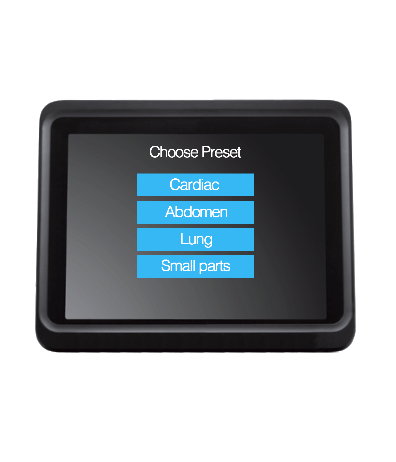
Three types of transducers are used in basic emergency ultrasound:
• Linear transducers
• Wide footprint curved abdominal transducers
• Smaller footprint cardiac transducers

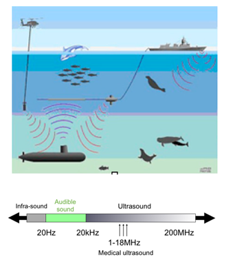
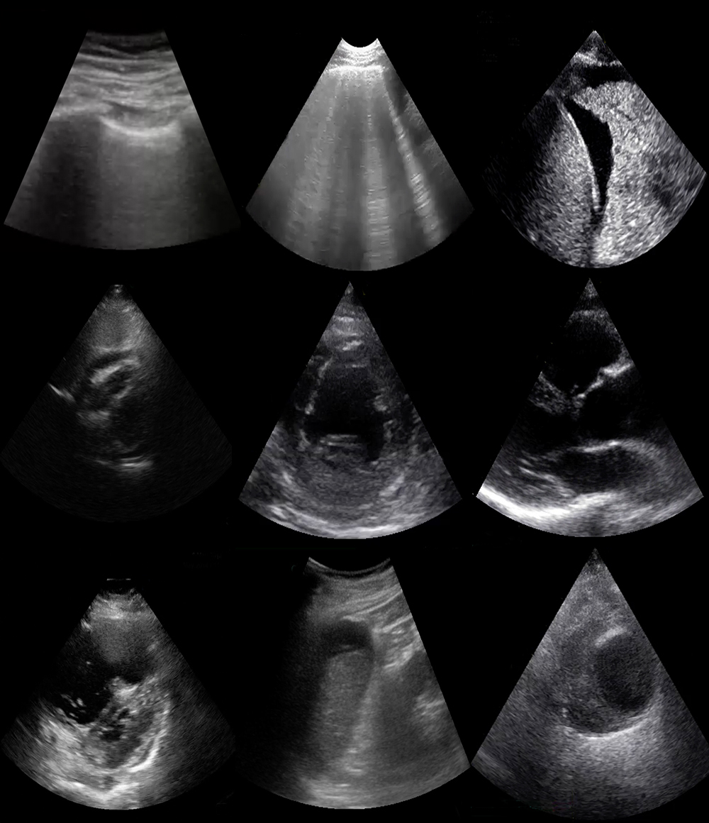
Transducer characteristics, such as frequency, determine the ultrasound image quality.

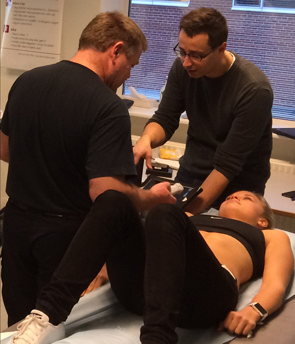
Linear transducers are high frequency transducers suitable for visualisation of superficial structures like the pleural line and peripheral vessels.

A low-frequency transducer like the curved abdominal transducer is suitable for better penetration of tissues in the abdominal and thoracic cavity.

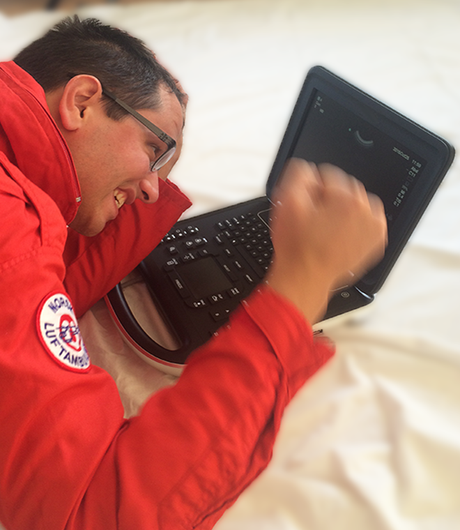
The cardiac transducers are especially designed to visualise the moving heart and are also suitable for assessing pleural fluid.

Image missing
Different ultrasound transducers – each made for a special purpose

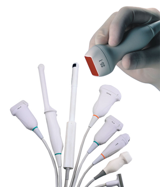
The hand is holding the preferred transducer type for cardiac examinations

Which preset?


The presets relevant for basic emergency ultrasound are:
• Lung
• Cardiac
• Abdomen

When possible, the appropriate preset for a given examination should always be selected.

In most ultrasound systems, the presets are chosen automatically when inserting or activating the relevant transducer.

A given preset for a given examination is not necessarily the optimal preset.

Different presets can be tried out to get the best image.

Image missing
Ultrasound system with preset menu open

Ultrasound made simple


The first look at an ultrasound machine can be an intimidating experience.

You see a bunch of unfamiliar buttons and knobs and confusion sets in. The user manual is often poorly designed, which only adds to the confusion. You turn it on, stare at a screen, and feel lost.

But, it can be made simple and easy, and you can quickly master the machinery through a basic understanding of ultrasound and by using a few controls.

Learn the few essential buttons – On/Off, select transducer, select preset, adjust gain and depth – and you are up and running.

Image missing
Cannot find the right buttons ….. aaaargh !!!

What is ultrasound?


Physics
In physics, the word “ultrasound” refers to sound with a frequency the human ear cannot perceive.

Application
Ultrasound is used among other things as a sonic depth finder in navigation, for cleansing of sensitive objects (e.g. contact lenses) and in testing non-disruptive materials in order to detect faults and fractures.

In medical diagnostics, ultrasound can be used for visualisation of anatomical structures and for assessment of physiological processes.

The ultrasound used for medical diagnostics is usually between 2 and 18 MHz.

Image missing

Course objectives


By completing this course you will learn:

• How to perform the focused ultrasonography examinations that are essential in emergency care
• What to look for
• How to answer nine focused questions that make a difference in emergency care

We will not teach you when to perform focused ultrasonography – this depends on you, the patient and the setting.

Image missing
Ultrasonography course

Please provide feedback


USabcd always strives to improve the quality of the
e-learning. We need your help to do this.

Therefore, any feedback provided by you is very important.

After each lesson there will be an opportunity for you to give your feedback regarding the content of the slide.

Feel free to give us any feedback you may have – comments, suggestions, praise, criticism, error reports etc.

The focused questions


The focused questions selected for this course are:

Heart
Is pericardial fluid collection present?
• Is the left ventricular function reduced?
• Are signs of pulmonary embolism present?
• Are signs of ascending aortic dissection present?

Lung
• Can pneumothorax be confirmed or excluded?
• Are signs of pulmonary edema present or can pulmonary edema be excluded?
• Can pleural fluid be confirmed or excluded?

Abdomen
• Is an abdominal aortic aneurism present?
• Is free intraperitoneal fluid visible?

These focused questions are not static – questions can be added and removed according to evidence and to the development of the ultrasound equipment.

Image missing
Demonstration of how the 9 pathologies presented in this course is seen on the ultrasound screen

What is Basic POCUS ?


Basic POCUS is ultrasonography in every emergency care provider’s pocket
This course teaches you how to use an ultrasound transducer like you use your stethoscope.

The content has been carefully selected to answer the questions on life-threatening and time-critical conditions, focused ultrasonography can answer.

Nothing is left out because of complexity – but everything has been boiled down to the bare essentials of focused ultrasonography in an emergency care setting.

There are no algorithms and no protocol names – the focus is solely on decision-making in individual patients using ultrasonography.

Image missing
Ultrasonography in the pocket

Focused ultrasonography


Concept
In this Basic POCUS course, the concept is focused ultrasonography.

Compared to protocolized ultrasonography, it represents a paradigm shift as it applies specific parts of the systematic ultrasonography examinations to solve specific clinical issues.

Only the necessary examinations are performed – and they depend on you and the clinical question you ask regarding your patient.

Image missing
Focused ultrasonography can be perfomed by everyone, everywhere and anytime.
The picture shows focused ultrasonography perfomed at the top of Kilimanjaro (5895 moh), performed by a medium trained physian with light hypoxia (SaO2 = 78%).

Focused questions criteria


The focused questions you ask must be of absolute importance to the patient in an emergency setting in terms of:

• Treatment
• Triage
• Transport method/preparation
• Qualifications of the receiving emergency care team

Whether a certain examination is of crucial importance or not depends on the situation, and the decision should always be weighed against the time required to do the examination.

Image missing
Focused ultrasonography performed in a patient’s home.