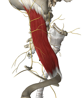
A lateral view of the point of emergence of the nerves of the superficial cervical plexus at the postero-lateral margin of the sternocleidomastoid muscle, where the nerves pierce the superficial cervical fascia to become subcutaneous

A lateral view of the point of emergence of the nerves of the superficial cervical plexus at the postero-lateral margin of the sternocleidomastoid muscle, where the nerves pierce the superficial cervical fascia to become subcutaneous
Place the patient supine with the head turned slightly contralateral
Use in-plane approach with the high-frequency linear probe in the axial plane at the level of the C4 nerve root typically corresponding to the level of the carotid bifurcation; alternatively the transverse process of C7 can be identified (the only transverse process with no anterior tubercle) and count the transverse processes upwards to the transverse process of C4
At the level of the C4 transverse process, the needle is inserted into the fascial plane between the deep and superficial cervical fascia deep to sternocleidomastoid muscle and from the postero-lateral margin of the sternocleidomastoid muscle with in-plane technique
Inject 20 mL of local anaesthetic to fill up the fascial space between the two cervical fascia

Tran DQH et al.: Analgesia for clavicular fracture and surgery. A call for evidence. Regional Anesthesia and Pain Medicine 38(6): 539-543 (2013)

A deep cervical plexus block blocks both the deep and superficial branches of the cervical plexus. It is a paravertebral block of the spinal nerve roots C2-C4
The indications are:
– carotid endarterectomy
– excision of cervical lymph nodes
The deep cervical plexus block anaesthetises both the sensory and the motor nerve branches of the cervical plexus, while the superficial cervical plexus block anaesthetises the sensory branches solely
Because the sensory blockade is equivalent for superficial and deep cervical plexus blocks, and because the deep block is more risky, the superficial cervical plexus block is generally favoured compared to the deep cervical plexus block

The innervation of the clavicle is controversial (ref. Tran et al, 2013)
Apparently, the supero-anterior aspect of the clavicle is innervated by the supraclavicular nerve from the superficial cervical plexus while the other parts of the clavicle are innervated by nerve branches from the brachial plexus: the suprascapular nerve and the long thoracic nerve (both from the superior trunk of the brachial plexus) – and maybe also the subclavian nerve
The sclerotomal innervation of the clavicle is traditionally ascribed to the C4 spinal nerve root
In 2/3 of human beings the C4 root contributes to the upper trunk of the brachial plexus; or maybe the sclerotomal innervation to the clavicle comes from the C4-C6 nerve roots

Supraclavicular nerve (from C3-C4 nerve roots) innervates the skin above and below the clavicle via the cervical plexus
Long thoracic nerve (from C5-C7 nerve roots) innervates the serratus anterior muscle via the brachial plexus
Subclavian nerve (from the superior trunk of the brachial plexus) innervates the subclavian muscle
Suprascapular nerve (from the superior trunk of the brachial plexus) innervates the supra- and infraspinatus muscles

Lesser occipital nerve (C2 nerve root) innervates the superior posterior cutaneous surface of the auricle and the cutaneous surface of the mastoid
Great auricular nerve (C2-C3 nerve roots) innervates the skin overlying the parotid gland, the external acoustic meatus, and the lower surface of the ear
Transverse cervical nerve (C2-C3 nerve roots) innervates the anterior and lateral aspects of the neck with ascending and descending branches
Supraclavicular nerve (C3-C4 nerve roots) innervates the skin above and below the clavicle, the sternoclavicular joint (medial branch), and the acromioclavicular joint (lateral branch)

The superficial cervical fascia envelopes the sternocleidomastoid muscle. The nerve branches of the superficial cervical plexus pierce the superficial cervical fascia to become subcutaneous
The deep cervical fascia (synonym: the prevertebral fascia) contains all the deep cervical muscles and all the branches of the deep cervical plexus
The nerve branches of the superficial cervical plexus run in the fascial plane between the deep and superficial cervical fascia before they become subcutaneous

Superficial cervical plexus block is indicated for head and neck analgesia:
– Central venous cannulation
– Anterior neck surgery
– Thyroid surgery
– Shoulder surgery “cape of anaesthesia”
– Carotid endarterectomy (CEA) – the carotid artery is innervated by the vagus and glossopharyngeal nerves as well as sympathetic branches from the superior cervical ganglion which requires local anaesthetic injected in the carotid sinus region for surgical anaesthesia
– Clavicular fracture – combine superficial cervical plexus and brachial plexus blocks