UGRA

13 – Tracking each spinal nerve root to the sulcus of the spinal nerve of the transverse process

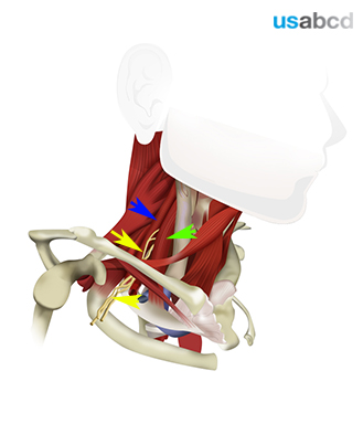
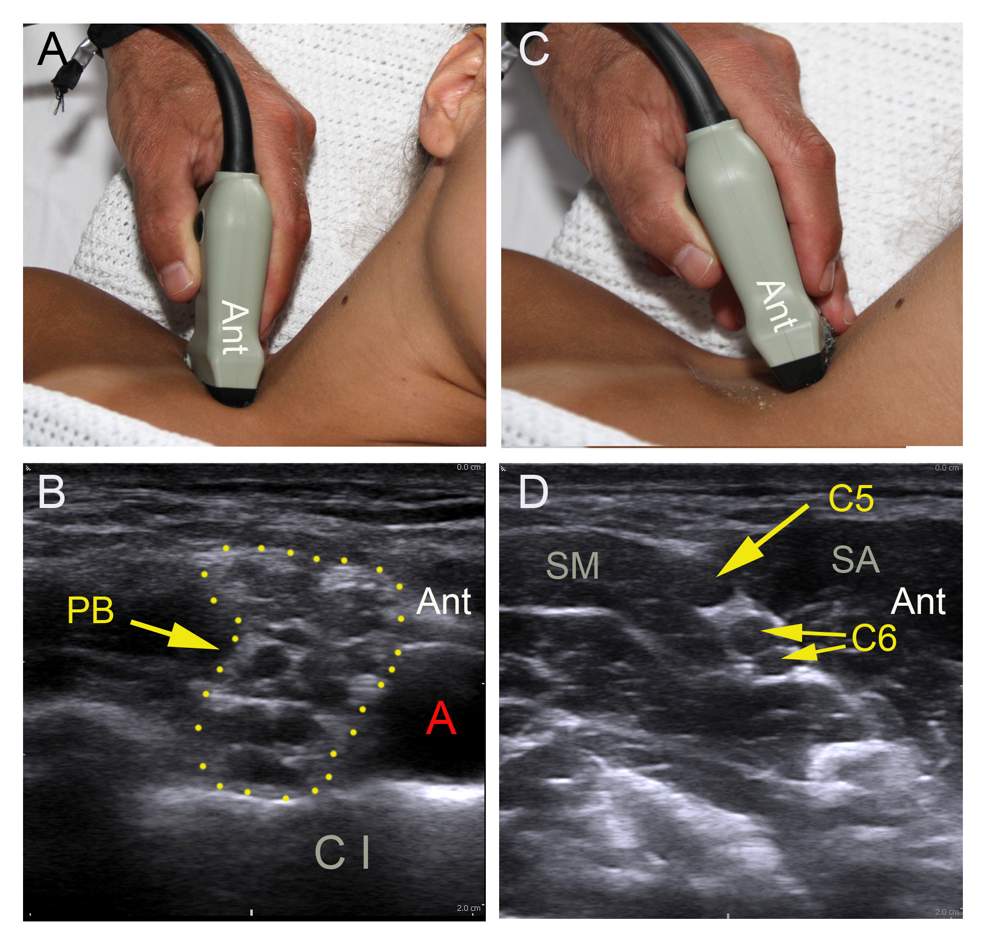
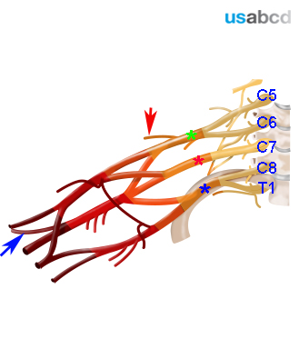
Each spinal nerve can be tracked from the branches of the brachial plexus in the transition zone behind the subclavian artery on top of the first rib to the sulcus of the spinal nerve of each transverse process

Click on the VIDEO CLIP button to view the video

Image missing
The video shows tracking of C8, C7, C6 and C5 to their sulci on the transverse processes
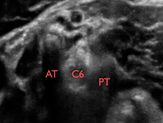
AT = anterior tubercle; PT = posterior tubercle; C5, C6, C7, C8, and T1 = the spinal nerve roots of the brachial plexus

3 – The muscle innervation of the shoulder

The shoulder muscles are innervated by

1) the axillary nerve (C5-C6; deltoid and teres minor)
2) the suprascapular nerve (C5-C6; supraspinatus and infraspinatus)
3) the subscapular nerve (C5-C6; subscapularis and teres major)
4) the medial and lateral pectoral nerves (C5-T1; major and minor pectorals)
5) the musculocutaneous nerve (C5-C7; coracobrachialis and long and short heads of biceps brachii)
6) the thoracodorsal nerve (C6-C8; latissimus dorsi)
7) the radial nerve (C5-T1; long and lateral heads of the triceps brachii)

The muscles innervated by the axillary and suprascapular nerves are the most clinically important for shoulder surgery

1)-3) and the major part of 4)-7) are anaesthetised by an interscalene brachial plexus block

Image missing
Some of the muscle innervation of the shoulder from an anterior and a posterior view. Deltoid (blue, axillary nerve), teres major and minor (grey, subscapular and axillary nerve), long and short heads of biceps brachii (yellow, musculocutaneous nerve), long and lateral heads of triceps brachii (orange, radial nerve).

4 – The cutaneous innervation of the shoulder

The cutaneous innervation of the shoulder is:

1) the axillary nerve (C5-C6; superior lateral brachial cutaneous nerve)

2) the supraclavicular nerve (C4; from the cervical plexus)

3) intercostal nerves (T2-T4; anterior cutaneous branches)

4) radial nerve (C5-T1; inferior lateral brachial cutaneous branches)

The interscalene brachial plexus block typically covers only C5-C6

Image missing
Innervation: Axillary nerve (cyan), supraclavicular nerve (pink), lateral cutaneous nerves (purple), medial brachial cutaneous nerve (green), inferior lateral brachial cutaneous nerve (from radial nerve, yellow).

5 – Anatomy of the brachial plexus in the interscalene groove

The brachial plexus is sandwiched between the anterior and the middle scalene muscles in the interscalene groove

Image missing
The figure shows the yellow branches of the brachial plexus (yellow arrows) emerging from between the anterior and middle scalene muscles
MSM = middle scalene muscle (blue arrow); ASM = anterior scalene muscle (green arrow)

8 – Indications of the interscalene block

The indications of the interscalene block are:

– Anaesthesia for shoulder surgery & shoulder
procedures

– Analgesia after shoulder surgery & shoulder
procedures

The ultrasound guided interscalene brachial plexus block is a basic level block

Image missing
A dislocated shoulder joint can be reduced using an interscalene brachial plexus block

9 – How to perform an ultrasound guided interscalene brachial plexus block

– Select a high-frequency linear probe
– Adjust gain, focus, and depth 2-3 cm
– Turn the orientation mark on the probe postero-lateral on the right side and antero-lateral on
the left side of the patient
– Place the probe behind the clavicle on top of the first rib oriented practically in the
parasagittal plane
– Locate the black, pulsatile subclavian artery (SA) on top of the first rib between the anterior
and the middle scalene muscles
– Locate the branches of the brachial plexus (BP) posterior to the SA
– Track the BP branches proximally until the profiles of the C5 and C6 spinal nerve roots line
up like a string of black pearls in the interscalene groove. C5 appears as one profile, C6 as
two
– Capture the best possible cross sectional image by tilting the probe and fine-tuning gain,
focus, and depth

Image missing
(A) Starting position (B) subclavian artery (A), costa I (CI) and brachial plexus branches (PB) (C) Track the brachial plexus branches proximally (D) C5 and C6 spinal nerve branches into the intescalene groove where they are sandwiched between the anterior scalene (SA) and the middle scalene (SM)

2 – The bone innervation of the shoulder

The shoulder and the shoulder joint is innervated by the axillary nerve and the suprascapular nerve

Minor contributions from the long thoracic nerve and capsular filaments from the infraspinatus nerve are not clinically significant

Image missing
The figure shows the bony structures of the shoulder from an anterior and a posterior view. The axillary nerve innervates the anterior side (purple). The suprascapular nerve innervates the posterior side (“bone” color). The blue color is innervated by the radial nerve.

1 – Introduction to Basic UGRA (Ultrasound Guided Regional Anaesthesia)

Basic UGRA (Ultrasound Guided Regional Anaesthesia) consists of easily learned and safe peripheral nerve blocks allowing effective perioperative analgesia for the majority of surgical procedures involving the upper and lower limbs and the anterolateral abdominal wall

The basic nerve blocks are exclusively performed as single shot injections using the in-plane needle approach to the target nerve

Image missing
The in-plane needle approach to the tibial nerve (sciatic nerve branch) in the popliteal fossa

2 – The learning objectives

The learning objectives of the basic ultrasound guided regional anaesthesia program are:

– Understanding the basic anatomy and sonoanatomy of the basic nerve blocks

– Understanding how to perform each of the basic nerve blocks

– Knowledge about the pitfalls

Image missing
The intercostal TAP block: local anaesthetic is injected between the posterior rectus sheath and the transversus abdominis muscle

1 – Anatomy of the brachial plexus

The brachial plexus (BP) innervates the upper limb

The BP originates from five spinal nerve roots: C5 to T1

The shoulder is innervated by the suprascapular nerve and the axillary nerve. They both originate from the spinal nerve roots C5 and C6

Anaesthesia of the shoulder requires blockade of the BP proximal to the level of the trunks, because the suprascapular nerve branches off the superior trunk

Image missing
The brachial plexus is displayed with the suprascapular nerve (red arrow) and the axillary nerve (blue arrow). The five spinal nerve roots of the brachial plexus are indicated by C5 to T1. Green asterix = superior trunk; red asterix = middle trunk; blue asterix = inferior trunk.