UGRA

2 – Bone distribution of the nerves from the brachial plexus

The infraclavicular brachial plexus block anaesthetize all the bones and joints of the upper limb distal to the shoulder:

– The axillary nerve innervates the head of the humerus

– The radial nerve innervates the arm, the elbow, the forearm, the hand and the
radial fingers

– The musculocutaneous nerve innervates the humerus

– The ulnar nerve innervates the elbow and the hand and the ulnar fingers

– The median nerve innervates the elbow and the forearm and the hand and the
radial fingers

Image missing
“Bone colour” = suprascapular nerve; green = axillary nerve; purple = radial nerve;
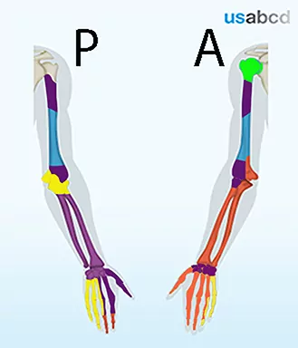
blue = musculocutaneous nerve; yellow = ulnar nerve; orange = median nerve

19 – Do not penetrate the brachial plexus nerves in the interscalene groove

It is important to advance the needle tangential to the target nerves and NOT penetrate the nerves

Click on the VIDEO CLIP button to view the video

Image missing
By advancing the needle tangentially to the nerves, it is possible to avoid piercing the nerves
C5 = spinal nerve root of C5; C6 = spinal nerve root of C6; C7 = spinal nerve root of C7; ASM = anterior scalene muscle; MSM = middle scalene muscle

14 – The in-plane approach to the interscalene brachial plexus block

The in-plane approach to the interscalene brachial plexus block

Local anaesthetic is deposited lateral to C5 and C6

You often feel a “pop” as you enter the interscalene groove

Click on the VIDEO CLIP button to view the video

Image missing
The video shows the in-plane approach to the interscalene block of the brachial plexus
ASM = anterior scalene muscle; MSM = middle scalene muscle; C5 = spinal nerve root of C5; C6 = spinal nerve root of C6

15 – Tracking the brachial plexus from the subclavian artery to the interscalene groove

To localise the brachial plexus you should start by localising the pulsating subclavian artery where it runs across the first rib

The branches of the brachial plexus are located just lateral to the artery

Track the branches of the brachial plexus proximally until they line up as a string of black pearls in the interscalene groove

Click on the VIDEO CLIP button to view the video

Image missing
The video shows the tracking of the branches of the brachial plexus from the level of the subclavian artery to the interscalene groove
SA = subclavian artery; BP = cluster of branches of the brachial plexus; ASM = anterior scalene muscle; MSM = middle scalene muscle

16 – Relocating the needle tip

If the perineural spread of local anaesthetic is not perfect, the needle can be relocated in real-time before injection of the remaining local anaesthetic

Click on the VIDEO CLIP button to view the video

Image missing
Relocate the needle tip if necessary to obtain perineural spread of local anaesthetic
C5 = spinal nerve root of C5; C6 = spinal nerve root of C6 (2 profiles); ASM = anterior scalene muscle; MSM = middle scalene muscle

17 – Relocating the needle tip – continued

Another example of relocation of the needle tip to obtain the endpoint of spread of local anaesthetic alongside the spinal nerve branches of C5 and C6

Click on the VIDEO CLIP button to view the video

Image missing
The video demonstrates relocation of the needle tip in order to cover the C5 and C6 spinal nerve branches with local anaesthetic in the interscalene groove
C5 = spinal nerve root of C5; C6 = spinal nerve root of C6; C7 = spinal nerve root of C7; A = small artery

18 – The contrast effect of the local anaesthetic

When local anaesthetic is injected it generates a contrast effect that enhances visualisation of the target nerves, because of the black background of the fluid

Click on the VIDEO CLIP button to view the video

Image missing
Bright are the stars that shine. Dark is the sky
C5 = spinal nerve root of C5; C6 = spinal nerve root of C6; LA = local anaesthetic

10 – The in-plane approach to the interscalene brachial plexus block

– Insert the needle from the posterior end of the probe

– Empty the hose and the needle of air by injecting 1/2 mL of local anaesthetic subcutaneously before advancing the needle

– Advance the needle with in-plane technique until the needle tip touches the lateral side of the BP between C5 and C6

– The endpoint of injection is complete spread of local anaesthetic along the lateral side of the C5 and C6 spinal nerve branches

– Reposition the needle tip as necessary to reach the endpoint

Image missing
The in-plane approach to the interscalene brachial plexus block
The in-plane approach is recommended as the first choice, as it allows real-time control of the position of the needle tip at all times

11 – String of black pearls

At the top of the interscalene groove the brachial plexus presents as the C5 and the C6 spinal nerve branches – and usually the C6 is split in two branches

The three profiles of C5 and C6 are usually lined up like a string of black pearls sandwiched between the anterior scalene muscle and the middle scalene muscle

Image missing
The string of black pearls made up by the spinal nerve branches of C5 and C6

12 – The spinal nerve roots can be tracked to the sulcus of the transverse process

It is possible to follow the spinal nerve roots all the way to the sulcus for the spinal nerve of the transverse process of the appertaining cervical vertebra

The seventh cervical vertebra does not have an anterior tubercle

Image missing
A drawing of the fourth cervical vertebra showing the sulcus for spinal nerve at the end of the transverse process between the anterior tubercle and the posterior tubercle