Local anaesthetic is injected just lateral to the femoral artery in the triangular space between the femoral artery, the sartorius muscle and the medial vastus muscle
Click on the VIDEO CLIP button to view the video
The video shows the in-plane approach to the saphenous nerve block with injection of local anaesthetic on the lateral side of the femoral artery SN = saphenous nerve; SM = sartorius muscle; FA = femoral artery; MVM = medial vastus muscle
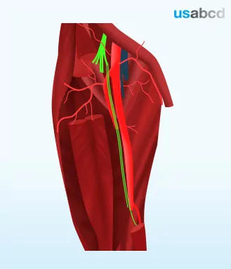
The saphenous nerve is the biggest branch from the femoral nerve
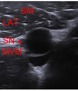
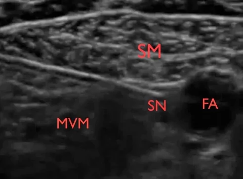
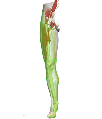
The saphenous nerve runs in the thigh in the femoral triangle and the adductor canal alongside the femoral vessels. In the femoral triangle and the upper part of the adductor canal the saphenous nerve is lateral to the vessels
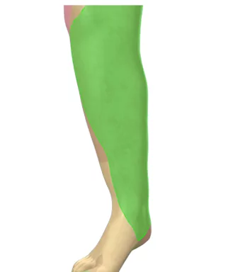
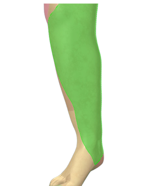
The saphenous nerve is 100% sensitive innervating the medial part of the knee capsule, the skin and subcutaneous tissue antero-inferior to the knee, of the medial leg, the medial ankle joint and the subtalar joints
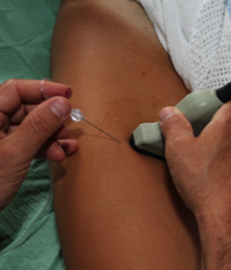
Indications: For surgical anaesthesia or pain management of the leg you should combine a sciatic and a saphenous nerve block
The figure shows the green saphenous nerve running alongside and lateral to the femoral artery in the femoral triangle
In the femoral triangle and the adductor canal the femoral artery is located under the triangular sartorius muscle (SM)
The saphenous nerve (SN) is sandwiched between the femoral artery (FA), the SM and the medial vastus muscle (MVM) in the femoral triangle and the proximal end of the adductor canal (AC). In the distal end of the canal the nerve is usually medial to the artery
Use in-plane (IP) approach from the lateral end of the transducer in the midthigh midways between the anterior superior iliac spine and the base of patella which is inside the femoral triangle
Complete perineural spread of local anaesthetic usually requires only a few milliliters of local anaesthetic and the duration of anaesthesia is oftentimes very prolonged – often more than 15 hours with ropivacaine
The image shows the FA underneath the SM in the AC. Often, the SN nerve is not distinctly visible prior to injection of local anaesthetic. However, in the upper half of the thigh the nerve is invariably located just lateral to the artery between the SM and the MVM. Lat = lateral side of the patient
The saphenous nerve is a branch of the posterior branch of the femoral nerve
The saphenous nerve innervates the antero-medial part of the knee and the leg including the medial malleolus and and the anterior side of the ankle joint and the subtalar joints
The green colour depicts the cutaneous distribution of the saphenous nerve
The anterior branch of the femoral nerve gives off the intermediate and the medial cutaneous nerves of the thigh
– the intermediate cutaneous nerve of the thigh innervates the skin of the anterior thigh
– the medial cutaneous nerve of the thigh innervates the skin of the medial thigh and knee
The posterior branch of the femoral nerve gives off the saphenous nerve, which is a sensory nerve innervating the antero-medial parts of the knee and the leg – sometimes as far distal as the big toe
The green shaded area depicts the cutaneous distribution of the femoral nerve branches
– the hip joint – the shaft of the femoral bone – the anterior thigh – the knee joint – the medial leg including the medial and anterior side of the ankle joint and the subtalar joints
The ultrasound guided femoral nerve block is a basic level block
The image displays the triangular hyperechoic femoral nerve (FN) in short-axis view just lateral to the femoral artery (FA). The FN is located on top of the iliopsoas muscle (IPM)
– Place a linear high-frequency probe in the inguinal groove and identify the pulsatile femoral artery – Align the artery to the centre of the monitor by adjusting the position of the probe and the depth of the image – Locate the femoral nerve (FN) lateral to the artery on top of the iliac muscle. The FN is usually flattened, spindle-shaped or triangular – rarely round – Capture the best possible cross-sectional image of the FN by tilting the probe 10-30 degrees cranially – Insert the needle from the lateral end of the probe – Advance the needle inside the plane of the ultrasound beam (in-plane, IP) until the tip of the needle touches the deep surface of the FN tangentially – The endpoint is complete perineural spread of local anaesthetic
Yellow arrowheads point at the femoral nerve; A is the femoral artery; IB is the inguinal boundary; LAT = lateral side of the patient; MED = medial; POST = posterior side of the patient