EEU

Transverse pelvic view – identifying structures Copy BUA


The pelvic view is one of the most difficult to interpret as a novice.

This is because of a lot of shadowing and disturbance from the surrounding structures e.g. bowel and bony structures of the pelvis.

In the image below this is especially seen as the dark areas of bony shadowing on each of the sides of the male bladder in the male pelvic view.

These areas should not be confused with intraperitoneal fluid.

Image missing
Female:
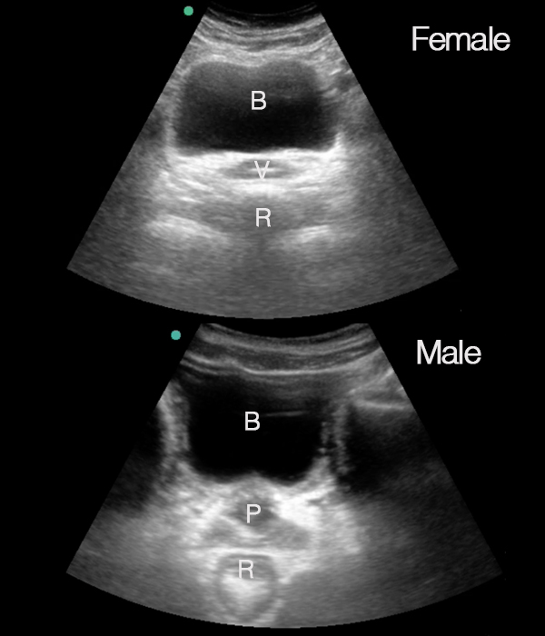
B: Bladder, V: Vagina, R: Rectum
Male:
B: Bladder, P: Prostate, R: Rectum

Perisplenic view: Identifying structures Copy BUA


In the perisplenic view, the following structures are seen:

• Diaphragm
• Spleen
• Kidney

Image missing
The image shows the scanning plane and the structures visualized in the perisplenic view.
D: Diaphragm
S: Spleen
K: Kidney
The splenorenal recess is marked in blue.

Obtaining the perisplenic view Copy BUA


The perisplenic view is obtained in the same way as perihepatic view.

Be aware that a slight rotation on the transducer in order to avoid rib shadowing should be done with a clockwise rotation as opposed to the counter-clockwise rotation in the perihepatic view.

Image missing
Perisplenic view

Perihepatic view: Identifying structures Copy BUA


In the perihepatic view, the following structures are seen:

• Diaphragm
• Liver
• Kidney

Image missing
The image shows the scanning plane and the structures visualised in the perihepatic view.
D: Diapgragm, L: Liver, K: Kidney. Morison’s pouch is marked in blue

Abdominal views when looking for free fluid Copy BUA


To look for free fluid in the abdomen, four views are relevant:

• The perihepatic view
• The perisplenic view
• The pelvic views – transverse and longitudinal views
• The peri-intestinal view(s)

The next sections will take you through how to obtain these views

Image missing
The image shows the relevant anatomical structures for assessing free intraperitoneal fluid.
Perihepatic view: Diaphragm, liver and kidney
Perisplenic view: Diaphragm, spleen and kidney
Pelvic views: Bladder and rectum – and in women also uterus (not demonstrated)
Peri-intestinal view

Slettes Abdominal aorta: Identifying structures Copy BUA


In the transverse aortic view, the following structures are seen:
• Aorta
• Inferior vena cava
• Columna vertebralis

In the longitudinal abdomnial aortic view, the following structures are seen:
• Aorta
• Liver (in the upper part of the abdomen)

Image missing

The transducer for focused ultrasonography of the abdomen Copy BUA


For focused ultrasonography of the abdomen a lower frequency curved (abdominal) transducer is used.

A lower-frequency curved transducer (abdominal) permits the deep penetration that is needed for evaluating deeper structures and has a wide footprint that allows a good overview.

If no abdominal transducer is available, a cardiac transducer can be used, but the small footprint of this transducer does not offer as good an overview.

Tips and tricks
The ‘abdominal’ transducer can be used for all lung and abdominal examinations.

Image missing
Curved abdominal transducer