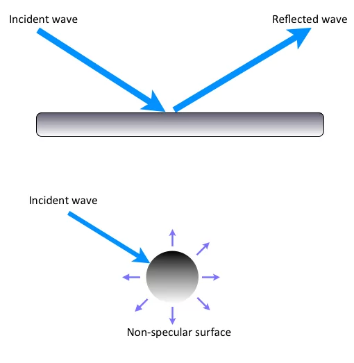
Medical student

Transmission and penetration

Transmission
When the sound wave propagates in a medium without striking any interfaces, it passes through the interface without any reflection, scattering or refraction (change of sound wave’s direction)

In a homogeneous medium the transmission is only reduced by absorption
Even in a heterogeneous medium some of the sound wave is usually transmitted when it strikes an interface

Penetration
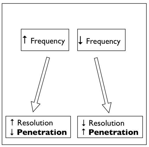
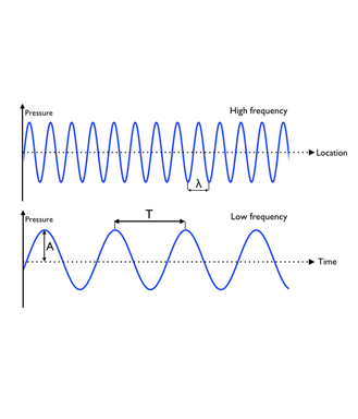
The ability of a sound wave to penetrate through tissue depends on the attenuation. This loss of penetration capacity is proportional to frequency

Penetration expresses how deep the ultrasound wave can penetrate down into the tissue

Image missing

Reflection

Ultrasound imaging is based on the reflection of sound, which are detectable echoes of the transmitted pulse

Reflection is a result of acoustic impedance mismatch

Reflection attenuates the sound wave reducing transmission beyond the reflecting interface. Reflected sound waves are synonymous to echoes – Reflection can be specular or diffuse

Specular reflection happens when the sound wave strikes a smooth surface (a specular reflector). In that case, the angle of incidence equals the angle of reflection

Diffuse (non-specular) reflection scatters the sound in multiple random directions. Diffusion occurs when the sound wave strikes small and irregular objects like red blood cells or interfaces in the tissue

In a completely homogeneous medium or tissue no reflection is made, and therefore no echoes are produced

Image missing
The figure shows specular and diffuse reflection.

What is ultrasound ?

Ultrasound is defined as sound with frequencies above the upper limit of the human hearing range of 20 kHz and up to 10 GHz

Its primary clinical application today is as a diagnostic tool and as a means to display anatomical structures, for which frequencies between 1 and 20 MHz are most commonly used

Image missing

XX Biological effect and possible damage

To date there have been no indications that the clinical use of ultrasound can compromise health

Ultrasound waves with an energy value below 100 W/cm2 do not cause significant tissue warming

100 W/cm2 is a limit that is not usually transcended in routine B-mode diagnostic ultrasound

Some of the effects of ultrasound that have been shown under laboratory conditions, such as the disruption of cell membranes, cavitation and formation of free radicals have not been demonstrated in the human body

To our knowledge, diagnostic ultrasound does not represent a risk factor for tissue damage

Image missing
A fetus visualized with ultrasound

Equipment and skills

A basic understanding of the user controls in the ultrasound system is required to learn ultrasound

Before you start clinical ultrasound scanning it is important to become familiar with the probe (or transducer) and the user interface of the ultrasound system

In this lesson you will learn about the probe and the most important user control buttons allowing you to optimize the ultrasound image quality

Image missing

Preparation of the ultrasound (US) system

Correct preparation of the ultrasound system will facilitate US examination and US guided procedures

– Ensure power supply for the procedure – either by connecting to network voltage or sufficient battery capacity
– Turn on the ultrasound system
– Choose the right “program” for the procedure (e.g. cardiac, nerve etc)
– Adjust the depth setting in order to align the target to the center of the monitor
– Adjust the gain setting
– Set the focus point

Image missing

The ultrasound transducer

Transducer characteristics, such as frequency and shape of the probe, determine ultrasound image quality

4 tranducers are used in clinical ultrasound
– Linear probes
– Curved array probes (abdominal)
– Microconvex array transducers
– Phased array transducers (cardiac)

Linear probes have frequencies in the range 8-20 MHz provide excellent 2D images, but have limited penetration due to their high frequency

Curved array probes (abdominal) with frequencies between 2-5 MHz produce adequate 2D images and permit deep penetration

Phased array transducers (cardiac) are optimised for scanning between ribs and penetrate deeply

Image missing
Example of linear, curved and microconvex probes

What is a medium?

A medium is the substance or material that carries the wave

The medium carries the wave from one point in the medium to another point

The medium is not the wave and it doesn’t create the wave; it merely carries or transports the wave from its source to other locations

The medium is made of particles, which are temporarily displaced and then return to the point of origin without transport of particles

A sound wave is transmitted by the vibration of the molecules in a medium. The medium can be air, water, wood, or any other material

Image missing
When a stone is dropped into a pond, the water molecules are disturbed
The molecules start oscillating around their original position – they vibrate

Waves

A sound wave is a mechanical wave

Mechanical waves require a medium in order to transport their energy from one location to another

They cannot propagate in vacuum

Mechanical waves can be longitudinal or transverse

A sound wave is a longitudinal wave

In a longitudinal wave the oscillating disturbance is parallel to the direction of travel

Sound waves are always longitudinal waves: The air molecules vibrate in the same direction as the sound wave travels and form a series of compressions (high pressure) and rarefactions (low pressure), where the molecules are squeezed together and pulled apart respectively

Image missing
A vibrating tuning fork creates a longitudinal wave. As the tines of the fork vibrate back and forth, they push on neighboring air particles. The forward motion of a tine pushes air molecules horizontally to the right and the backward retraction of the tine creates a low-pressure area allowing the air particles to move back to the left.

Sound wave characteristics

Waves can be described mathematically with a location-amplitude-coordinate system and a time-amplitude-coordinate system with a sinus curve

The basic parameters of a harmonic wave are:
• Amplitude (A) = maximum oscillation
• Wave length (lambda) = distance from wave top to wave top
• Period (T) = time interval from one wave top at a well defined location until the next wave top gets to the same location
• Frequency (1/T) = number of wave tops per time unit. SI unit for frequency is Hertz (Hz) = s-1
• Velocity (v) = the speed of the wave = lambda/T

Image missing