Basic Ultrasound Physics

22 – What is ultrasound?

Ultrasound is sound

Ultrasound is defined as sound with frequencies above the upper limit of the human hearing range of 20 kHz and up to 10 GHz (1 Gigahertz (GHz) = 1000 Megahertz (MHz) = 1 billion Hz)

Hertz is the number of cycles per second. In other words it is a measure of frequency

The primary clinical application of ultrasound today is as a diagnostic tool and as a means to display anatomical structures, for which frequencies between 1 and 20 MHz are most commonly used

Image missing
A high-frequency linear transducer is used to guide an interscalene brachial plexus block

23 – Ultrasound imaging

Medical ultrasound imaging is based on the impulse-echo principle of generating and emitting short pulses of ultrasound that is reflected at tissue interfaces and subsequently recorded by the receiver. The reflected sound wave is essential for the production of the ultrasound images. Sound energy reflected back to an ultrasound transducer strikes the piezoelectric crystal and makes it vibrate and convert the sound energy to electrical voltage.

The ultrasound system processes the information and calculates:
• the depth of the reflected echo
• the amplitude (energy) of the reflected sound wave

Medical ultrasound exploits the piezoelectric effect: high frequency sound waves are emitted when piezoelectric crystals are exposed to electric current. This was discovered by the brothers Pierre and Jacques Curie in 1888.

Image missing
The figure shows the emission of sound from piezoelectrical crystals in the transducer and reflection of the signal at tissue interfaces depending on the difference in acoustic impedance.

24 – The history of ultrasound 1793-1912

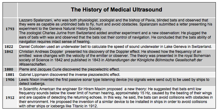
The figure shows the history of ultrasound in the period 1793-1912. The discovery that bats were using ultrasound for navigation, and the discovery of the piezoelectric crystal were the key findings in this period.

Image missing

25 – The history of ultrasound 1915-1995

The figure shows the history of ultrasound in the period 1915-1995 where the physics of ultrasound was established and the use of ultrasound in imaging was developed forming the premise for the use of ultrasound in medical imaging.

Image missing

26 – Biological effect and possible damage

Although energy is transmitted to the tissue during every ultrasound examination, to date there have been no indications that the clinical use of ultrasound can compromise health

As far as is currently known, ultrasound waves with an energy value below 100 W/cm2 do not cause significant tissue warming. This is a limit that is not usually transcended in routine B-mode diagnostic ultrasound

Some of the effects of ultrasound that have been shown under laboratory conditions, such as the disruption of cell membranes, cavitation and formation of free radicals have not been demonstrated in the human body

To our knowledge, diagnostic ultrasound does not represent a risk factor for tissue damage

Image missing
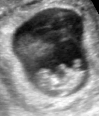
A fetus visualized with ultrasound.

29 – Summary

This chapter has explained the fundamental principles of wave and acoustic physics, and now you should be familiar with:

– waves, sound waves, and medium

– sound wave characteristics

– wave-medium interactions: attenuation, absorption, penetration, transmission,
acoustic impedance, reflection, refraction, and diffraction.

– ultrasound definition, imaging, and history

– biological effect and damage

In the next chapter the ultrasound system will be reviewed.

Image missing

16 – Acoustic impedance

Ultrasound is reflected when it crosses boundaries between different kinds of tissues. The reflection is caused by the change in impedance. Acoustic impedance describes how hard the sound wave has to push the tissue to make it move, or the resistance of the medium to vibration caused by a sound wave. The acoustic impedance is dependent on the density and sound velocity of the tissue.

Acoustic impedance is the only source of sound wave reflection. The importance of acoustic impedance is that a propagating sound wave is reflected only at interfaces between two tissue types with different acoustic impedances. This is known as impedance mismatch. The bigger the difference in acoustic impedance, the bigger is the reflection.

In ultrasound imaging strong reflection is displayed by white color (hyperechoic) on the screen. Many body tissues have similar acoustic impedances and only a small proportion of the sound is reflected at interfaces between two such media (e.g. fat, water, and soft tissues).
The opposite is seen when ultrasound is reflected at interfaces with different acoustic impedances.

Image missing
The table shows sound velocity and acoustic impedance in different tissues. When sound waves pass through a physical medium, the medium particles move due to the wave pressure. The medium’s acoustic impedance is the ratio of the sound wave pressure and the particle velocity it produces.

17 – Impedance mismatch, matching and ultrasound gel

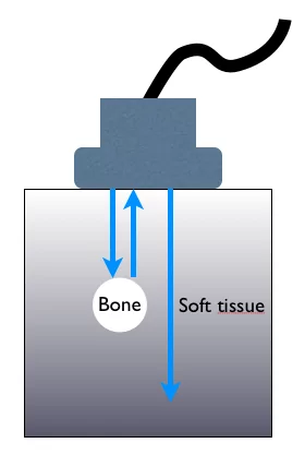
The acoustic impedances of air and bone are radically different from other tissues. As a consequence, interfaces between soft tissue and air (or bone) reflect most of the sound energy striking the interface. The practical implication is that ultrasound cannot be used to image structures located deep to bone or air interfaces.

Soft tissue and blood have slightly different acoustic impedances, which explains why the needle becomes more visible as it enters the blood vessel – the acoustic impedance difference between needle and blood is bigger than between needle and soft tissue.

The use of ultrasound gel is an example of impedance matching: The gel eliminates air between the transducer and the skin and creates a smooth acoustic impedance transition for the sound waves from probe to patient resulting in less reflected (wasted) energy.

Image missing
The figure shows the difference in reflection in bone and soft tissue, caused by the difference in the acoustic impedance in two tissues.

18 – Reflection

The creation of ultrasound images is based on the reflection of sound, which are detectable echoes of the transmitted pulse. Reflection is a result of acoustic impedance mismatch.

Reflection attenuates the sound wave reducing transmission beyond the reflecting interface. Reflected sound waves are synonymous with echoes.

Reflection can be specular or diffuse
Specular reflection happens when the sound wave strikes a smooth surface (a specular reflector). In that case, the angle of incidence equals the angle of reflection.

Diffuse (non-specular, scattering) reflection scatters the sound in multiple random directions. Scattering occurs when the sound wave strikes small and irregular objects or interfaces in the tissue.

In a completely homogeneous medium or tissue no reflection is made, and therefore no echoes are produced.

Image missing
The figure shows specular and diffuse reflection.

20 – Refraction and diffraction

Refraction
The sound wave is refracted (bent) when it is transmitted through an interface between two media. Usually the refraction is subtle. However, multiple refractions can add up to become significant.

The degree of bending depends on the change in propagation velocity between the medium on the incident side and the medium on the transmitted side (Snell’s Law).

Fat and bone cause significant refraction. Refraction can produce double image artefacts. If the velocity is greater in the first medium, the refraction occurs towards the perpendicular; if it is greater in the second medium, the refraction occurs away from the perpendicular.

Diffraction
The ultrasound beam diverges proportionally to distance from the transducer. The decrease of the intensity of the beam proportional to divergence is called diffraction.

Image missing
The figure displays the refraction of light at an interface between air and water due to the change in propagation velocity.