This includes:
– An ultrasound system suited for FLUS
– Reliable power – either external AC power or a battery
– Preset suited for FLUS
– Plenty of acoustic coupling agent – ultrasound gel
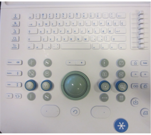
– Adjusting the image depth, image gain and focus
Image of an ultrasound machine keyboard; it is of vital importance to know where the depth, gain and focus buttons are placed
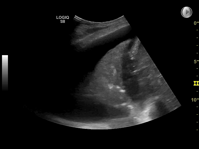
Microconvex array transducers with intermediate frequencies (7-4 MHz) produce good 2D images and acceptable ultrasound artefacts.
They are nicely shaped for scanning between the ribs in the intercostal spaces with good penetration in patients with a thick chest wall and are well suited for pleural evaluation.
Due to their small size, microconvex transducers are excellent for the assessment of the posterior chest surface of supine patients.
Transducer characteristics, such as frequency and shape of the probe, determine the ultrasound image quality.
Linear and curvilinear (or curved) transducers are most useful for FLUS imaging because they provide excellent image quality and produce the important air artefacts in FLUS (lung sliding, B-lines, and lung pulse).
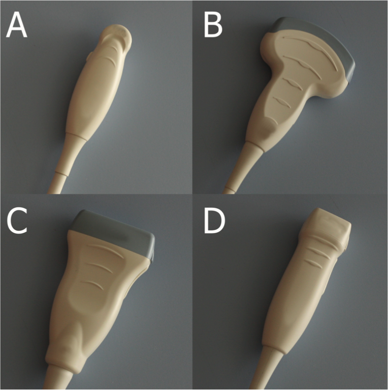
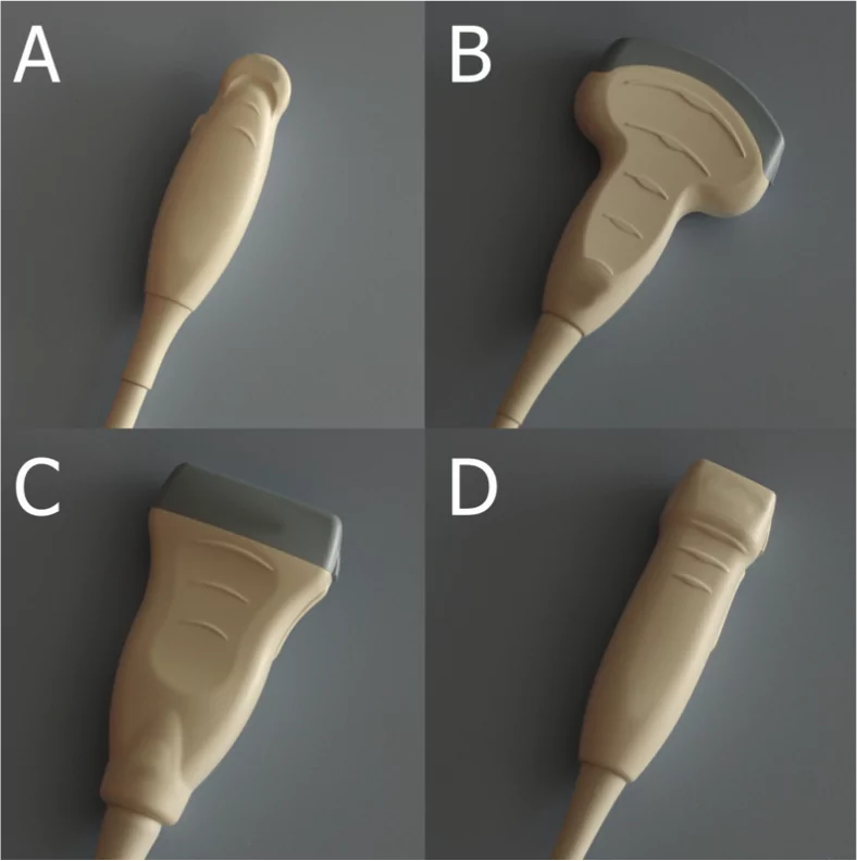
Different types of transducers:
A: Microconvex transducer, B: Curved array (abdominal) transducer, C: Linear transducer, D: Phased array transducer. A, B and C are suited for lung ultrasound.
Expensive newer generation ultrasound systems often contain sophisticated image smoothing software (e.g. harmonics, cross-beam) that is appropriate for some types of diagnostic ultrasonography.
The software is typically programmed to minimise the presence of artefacts, which are critically important in FLUS; knowledge on how to switch such software off and on is therefore necessary.
2D (also known as B-mode) and M-mode are sufficient for performing FLUS; more sophisticated scanning modes (e.g. colour Doppler mode) are generally not needed.
Not all ultrasound machines have a dedicated FLUS preset.
Additionally, some FLUS presets tend to be adjusted to diagnosing superficial pathology such as pneumothorax and are therefore not ideal for more profound pathology.
The operator needs to know which presets are available on the ultrasound machine and which presets generate the best images for a given clinical scenario.
If a FLUS preset is not available, a preset for an “abdominal examination” can often be used when diagnosing profound conditions such as pleural effusion.
A “superficial” or “musculoskeletal” preset can be used when diagnosing conditions solely located at the pleura.
The image demonstrates the keyboard of an ultrasound machine; choosing the correct presets is a key skill