By completing this course, you will gain the ability to:
Perform the focused ultrasound examinations pivotal in perioperative medicine.
Understand what to look for during these examinations.
Use ultrasound effectively to answer specific clinical questions relevant in a perioperative setting.
Note: While we’ll equip you with the how-tos, the decision of when to perform focused ultrasonography remains context-dependent, factoring in your judgement, the patient’s situation, and the specific setting.
Throughout this course, we’ll explore answers to critical questions that ultrasound can help address. These range from evaluating cardiac conditions to discerning lung and abdominal anomalies, ensuring you can make informed decisions in critical moments.
Key focused questions addressed in this course:
Heart:
Is there a pericardial fluid collection?
Is the left ventricular function reduced?
Are there indications of pulmonary embolism?
Signs of hypovolemia: What to look for?
Identifying hypertrophy: Key signs and indicators.
Lung:
Confirming or ruling out pneumothorax.
Detecting or excluding pulmonary edema.
Ascertaining the presence of pleural fluid.
Recognizing signs of atelectasis and consolidation.
Abdomen:
Identifying free intraperitoneal fluid.
Assessing the bladder: Is it full or empty?
Gastric ultrasound: Evaluating stomach contents to determine fullness, a crucial assessment prior to anesthesia to mitigate the risk of aspiration.
Demonstration of how 9 pathologies can be seen on the ultrasound screen.
Focused ultrasonography can be perfomed by everyone, everywhere and anytime.
The picture shows focused ultrasonography perfomed at the top of Kilimanjaro (5895 moh), performed by a medium trained physian with light hypoxia (SaO2 = 78%)
Point-of-Care Ultrasound (PoCUS) is the use of ultrasound at the bedside to either guide a procedure or answer a clinical question.
PoCUS is about leveraging ultrasound right at the patient’s bedside, either to guide a procedure or to answer a pressing clinical question. In this course, we train you to wield the ultrasound transducer for diagnostic purposes, honing in on the essentials to address foundational questions in perioperative medicine. We’ve distilled the content to its core, ensuring you master focused ultrasound in a perioperative context without any superfluous information.
Compared to protocolized ultrasonography, this course represents a paradigm shift as it applies specific parts of the systematic ultrasound examinations to solve specific clinical issues. There are no algorithms and no protocol names.
Only the necessary examinations are performed – and they depend on you and the clinical questions you ask regarding your patient.
You do not have to stick to a specific protocol!
The concept
– Ask a question
– If you can answer it with ultrasonography, pick up the ultrasound transducer
– When the question is answered, put down the transducer and take care of the patient
Basic UGRA (Ultrasound Guided Regional Anaesthesia) consists of easily learned and safe peripheral nerve blocks allowing effective perioperative analgesia for the majority of surgical procedures involving the upper and lower limbs and the anterolateral abdominal wall
The basic nerve blocks are exclusively performed as single shot injections using the in-plane needle approach to the target nerve
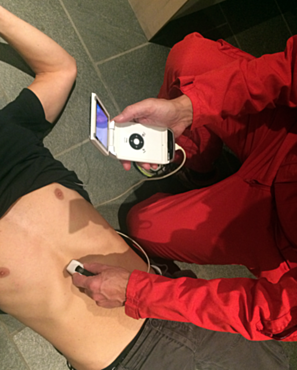
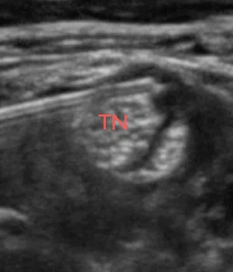
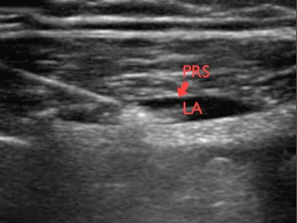
The in-plane needle approach to the tibial nerve (sciatic nerve branch) in the popliteal fossa
Basic UGRA (Ultrasound Guided Regional Anaesthesia) consists of easily learned and safe peripheral nerve blocks allowing effective perioperative analgesia for the majority of surgical procedures involving the upper and lower limbs and the anterolateral abdominal wall
The basic nerve blocks are exclusively performed as single shot injections using the in-plane needle approach to the target nerve
The in-plane needle approach to the tibial nerve (sciatic nerve branch) in the popliteal fossa
• How to perform the focused ultrasonography examinations that are essential in emergency care
• What to look for
• How to answer nine focused questions that make a difference in emergency care
We will not teach you when to perform focused ultrasonography – this depends on you, the patient and the setting.