Lung Ultrasound

15 – FLUS examination


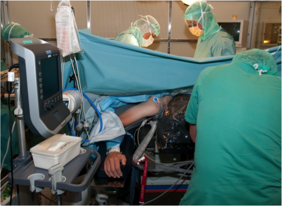
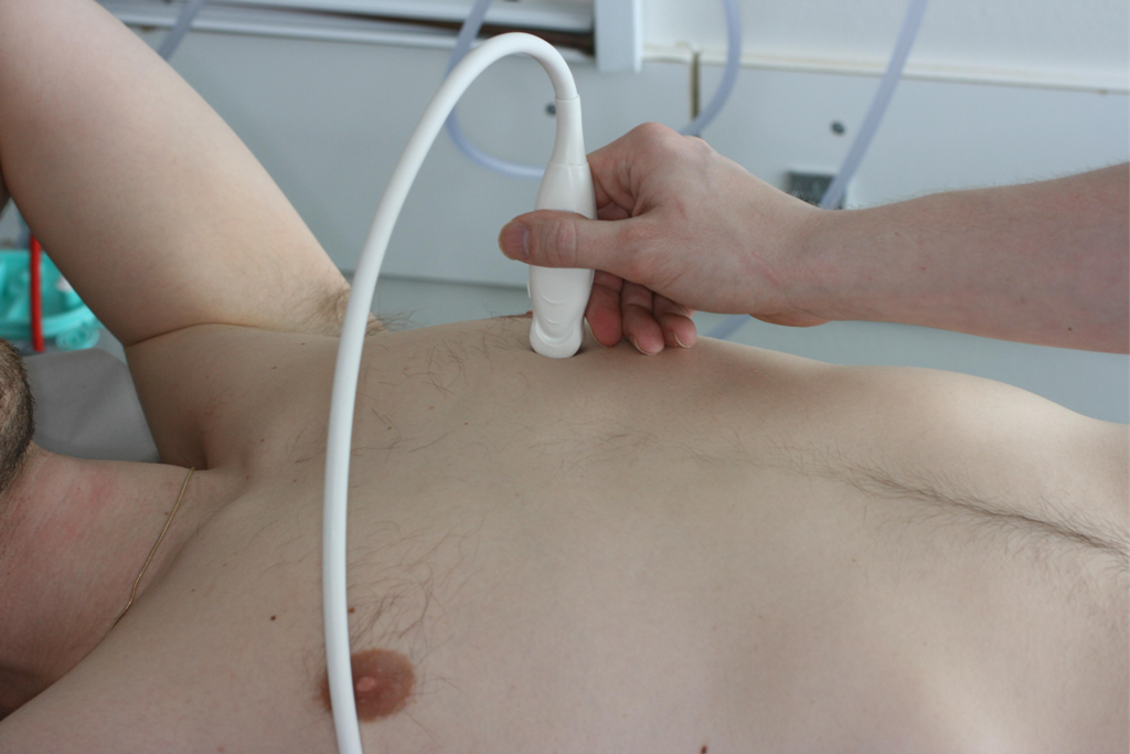
Since FLUS is used in critically ill patients in a variety of settings (e.g. prehospital, ED, ICU, OR) it may not always be possible to perform a standardised FLUS examination.

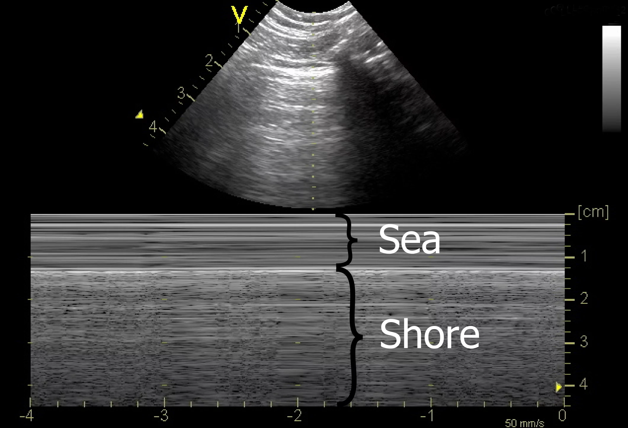
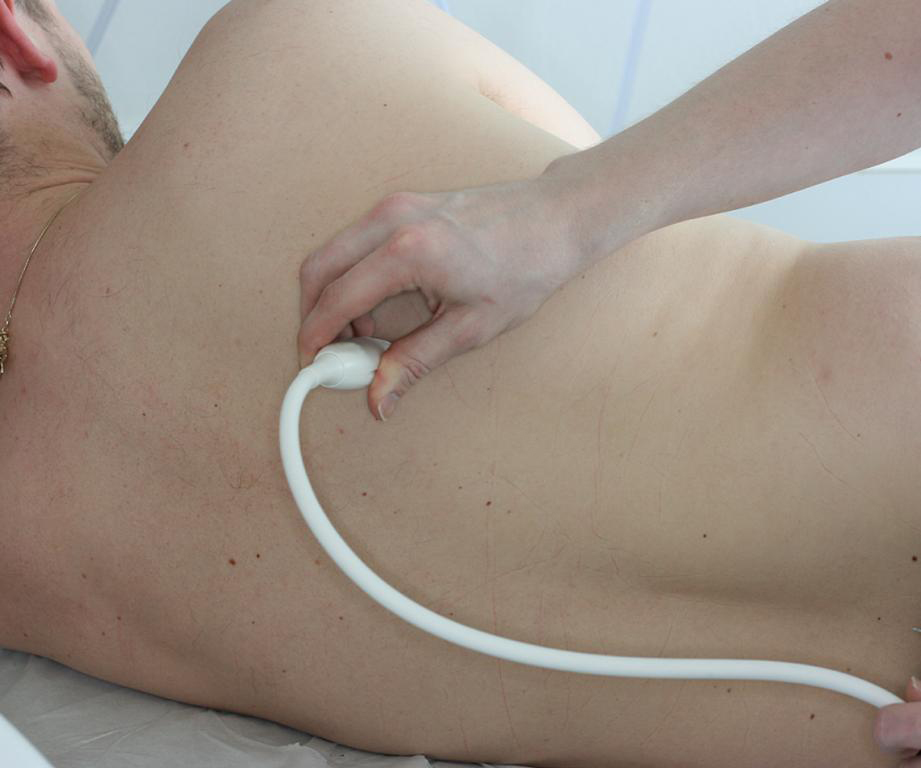
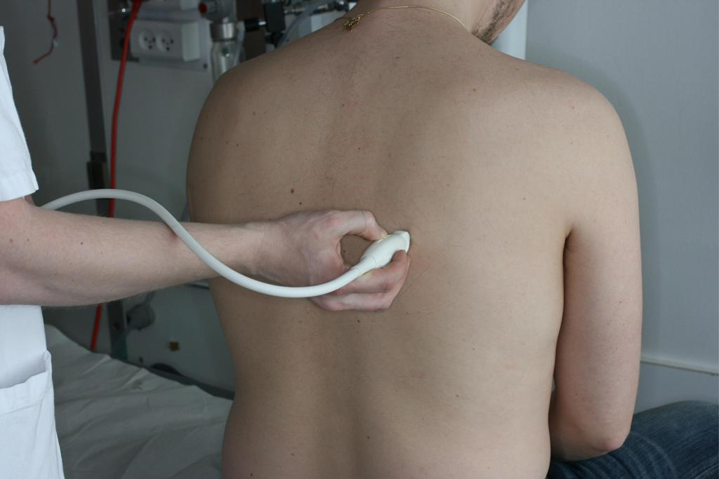
By using FLUS in a standardised and systematic approach, however, the examination still has a very high feasibility and the diagnosis and exclusion of important diagnoses such as pneumothorax, pleural effusion and interstitial syndrome can often still be made.

Image missing
FLUS can be a challenge in the OR.

14 – Ultrasound scanning modes


Normally B-mode is sufficient when performing FLUS.

Other modalities such as M-mode or colour Doppler can be used in some cases where there is doubt whether lung sliding is present or not (see lesson on pneumothorax).

Image missing
M-mode scanning of pleura with seashore sign.

13 – Patient position


Some patients with respiratory emergencies can not be placed in a supine position due to dyspnoea.

Anterior, lateral and posterior surfaces are then scanned with the patient in the sitting position.

In comparison, some critically ill patients may not be able to sit up for the assessment of the posterior zones.

The posterior surfaces can then either be scanned with the patient lying on the side or alternatively the transducer can be inserted in between the mattress and the patient making it possible to scan at least a part of the posterior surface.

Image missing
Image showing a microconvex transducer placed in zone 6R, with the patient lying on the side.

12 – FLUS examination technique continued


Once the anterior and lateral surfaces have been scanned, the patient is asked to sit up and the squares on the posterior surface are scanned using the same principles as described for the anterior and lateral surfaces.

Again in each scanning zone it is noted whether pneumothorax, pleural effusion or multiple B-lines are present or whether only normal findings are present.

Image missing
Image showing a microconvex transducer placed in zone 6R, with the patient in the sitting position.

11 – FLUS examination technique


With the patient in the supine position the transducer is quickly placed in each of the scanning squares.

The transducer is placed in a longitudinal axis over an intercostal space.

In each square it is noted whether pneumothorax, pleural effusion or multiple B-lines are present or whether only normal findings are present.

Image missing
Image showing a microconvex transducer placed in zone 2L.

28 – Summary

As with other kinds of sonography, FLUS findings cannot stand alone and should be used as an adjunct to clinical assessment and other diagnostic modalities

Presence or absence of the interstitial syndrome is a powerful tool with both a high sensitivity and specificity for ruling in and ruling out pulmonary edema in patients with acute dyspnoea

The interstitial syndrome has many causes why it is less specific in some settings such as a intensive care unit

Image missing
FLUS using a low-frequent abdominal transducer, multiple B-lines are present

24 – Acute respiratory distress syndrome (ARDS)

In ARDS the changes found with LUS are not necessarily gravity dependent

Hence, the number of B-lines are not necessarily most pronounced in the dependent part of the lungs

Furthermore, spared areas with no B-lines are often seen

As in interstitial lung disease, in ARDS the visceral pleura is also often affected, and therefore in areas can appear thickened and fragmented

Image missing
The FLUS clip is from zone 1R of a patient with ARDS

23 – Interstitial lung disease

Not all interstitial lung disease causes interstitial syndrome

In interstitial lung disease the pulmonary changes are not gravity dependent, hence the number of B-lines are not necessarily most pronounced in the dependent part of the lungs

Furthermore, spared areas with no B-lines are often seen

In interstitial lung disease, such as idiopathic pulmonary fibrosis, the visceral pleura is also affected

The visceral pleura therefore in some areas can appear thickened and fragmented which is often not the case in simple pulmonary edema

Image missing
The FLUS video is from the right lower posterior quadrant (5R) of a patient with idiopathic pulmonary fibrosis

22 – Causes of pulmonary edema

LUS cannot differentiate between cardiogenic and non-cardiogenic pulmonary edema

Supplementary focused assessed transthoracic echocardiography (FATE) is an ideal tool which can be used to assess whether interstitial syndrome is of cardiogenic origin or not

Image missing
The video demonstrates a FATE (subcostal view) in a patient who had interstitial syndrome due to decompensated systolic heart failure with pulmonary edema

21 – Pulmonary edema

In both cardiogenic and non-cardiogenic pulmonary edema the grade of increased density of the lungs are most severe on the posterior surface of the lungs compared to the anterior surface (if the patient has been lying down)

The number of B-lines tend to be higher more posteriorly than anteriorly and there are no sparred areas with no B-lines in the dependent regions of the lungs

The visceral pleura are not affected by pulmonary edema and the pleural line therefore appears normal.

The video is of LUS of the right upper anterior quadrant (1R) of a patient with cardiogenic pulmonary edema

Image missing
FLUS clip from a patient with cardiogenic pulmonary edema, multiple B-lines are present