LUSBas

17 – Differential diagnoses

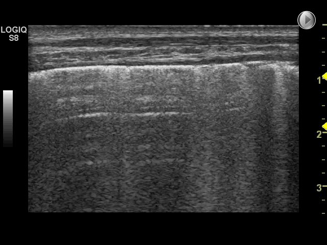
Based on patient history and primary assessment, a list of some of the most common and important differential diagnoses would be:

– COPD exacerbation
– Heart failure with pulmonary edema
– Pneumonia
– Pulmonary embolism
– Pleural effusion
– Pneumothorax

Image missing
Often only clinical assessment, ECG, ABGA and CXR can be performed quickly in the ED room

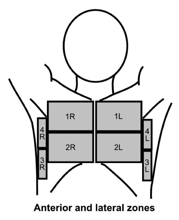
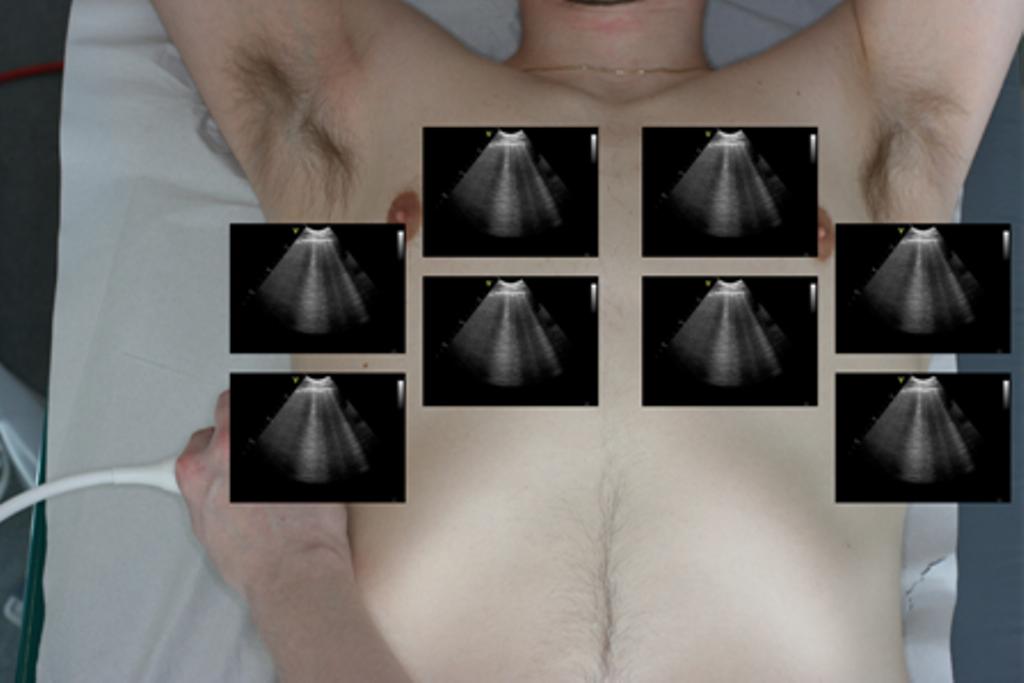
FLUS high feasibility and diagnostic capabilities warrants its use as a standard diagnostic test in patients with respiratory symptoms

16 – History and findings

68 year old female with severe COPD

Admitted with progressive dyspnoea and coughing, symptoms had lasted 14 days, possible fever

Primary assessment:
Auscultation: Prolonged expiration, wheezing. No murmurs. No edema or tenderness of the legs

Image missing

15 – Interstitial syndrome: Case

The following case illustrates how FLUS can be used in an emergency department to rule-in or rule-out cardiogenic pulmonary edema

Image missing
An US machine prepared and ready to perform FLUS in acute admitted patients

14 – Exclusion of the interstitial syndrome in COPD

Patients with exacerbation in chronic obstructive pulmonary disease (COPD) or asthma do not have interstitial syndrome

Hence, in acute admitted patients with dyspnoea, LUS can be used as a tool to differentiate between cardiogenic and pulmonary causes of dyspnoea

Image missing
The video is from a patient admitted with COPD exacerbation, the pattern is that of a normal FLUS

13 – The interstitial syndrome in Emergency patients

In an emergency department, the by far most common cause for interstitial syndrome is cardiogenic pulmonary edema

Hence, in this setting FLUS with signs of interstitial syndrome, one should suspect cardiogenic pulmonary edema

Image missing
FLUS is a key part of the initial assessment of patients with respiratory symptoms admitted to an ED

12 – The interstitial syndrome in ICU patients

In patients who have been admitted for a while in an intensive care unit (ICU), many diseases and conditions may cause interstitial syndrome

Hence, LUS with signs of interstitial syndrome, is a less specific finding in this setting

In patients with acute respiratory failure to an intensive care unit, the finding of interstitial syndrome is however most often due to cardiogenic pulmonary edema

Despite these limitations FLUS can still as an efficient tool to rule-out these conditions

Supplementary use of other kinds of point-of-care sonography such as focus assessed transthoracic echocardiography (FATE) can be of further use to differentiate between cardiogenic and non-cardiogenic causes of interstitial syndrome

Image missing
FLUS video from a patient with acute respiratory failure

Since no B-lines were present, IS was absent and pulmonary edema could be ruled-out

11 – Pulmonary edema

The finding of interstitial syndrome is a powerful diagnostic tool in diagnosis of pulmonary edema

FLUS is very sensitive for detecting interstitial oedema. Consequently, a FLUS examination with no signs of interstitial syndrome can be used to rule out conditions such as cardiogenic pulmonary oedema

Image missing
FLUS from zone 3R in a patient with cardiogenic pulmonary edema. Multiple B-lines and a small pleural effusion are present

10 – Causes of the interstitial syndrome

Any condition with universally increased density in the lungs may cause universally multiple B-lines and therefore the interstitial syndrome

In adults the following conditions have been described as causes of interstitial syndrome:

– Cardiogenic pulmonary edema
– Non-cardiogenic pulmonary edema (e.g. fluid overload, renal failure)
– Interstitial lung diseases
– Viral pneumonia
– Bacterial pneumonia
– ARDS
– Acute chest syndrome
– Drowning / near-drowning
– Lung contusion

Image missing
FLUS performed with a high frequency linear transducer

Multiple B-lines are present

9 – Criteria for the interstitial syndrome

Using the scanning technique described in the examination technique module, with two anterior, two lateral and three posterior scanning zones, the criteria for IS is the presence of both of the following:*

1: A positive scanning zone is defined by the presence of three or more B-lines in a longitudinal plane between two ribs

2: When scanning the anterior and lateral zones, two or more of the zones have to be positive on each side

* Volpicelli, G., et al., “International evidence-based recommendations for point-of-care lung ultrasound”, Intensive Care Med, 2012, 38(4): p. 577-91

Image missing
The image depicts the anterior and lateral zones used for the assessment of interstitial syndrome

8 – Definition of the interstitial syndrome

Multiple B-lines can in some diseases be seen almost universally when scanning both lungs. This finding is often referred to as the interstitial syndrome (IS)

Image missing
FLUS findings in a patient with multiple B-lines in all anterior and lateral zones bilaterally