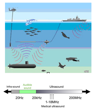
Lesson-UnderstandUS

Frequency, penetration and resolution


Frequency
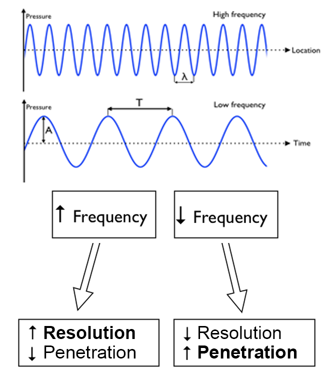
The frequency of a sound wave is equal to the number of wave tops per time unit.

The SI unit for frequency is Hertz (Hz) = wave tops pr second.

Ok, so that does not sound simple…

This is what you need to know:
The longer the distance the sound wave travels, the more energy is lost to absorption.

The ability of a sound wave to penetrate through tissue depends on the frequency of the sound wave – a lower frequency (lower number of wave tops) gives a better penetration.

On the other hand, a higher frequency (more wave tops) causes a higher resolution, but a lower penetration.

Image missing

Use of the ultrasound system


A systematic approach will facilitate successful scanning:

• Ensure a power supply for the procedure – either a connection to a network voltage, or sufficient battery capacity
• Turn on the ultrasound system
• Select your transducer
• Choose preset (predefined setting appropriate for a specific examination)
• Obtain view
• Adjust the depth setting
• Adjust the gain setting
• Interpret the findings

Image missing
Different point-of-care ultrasound systems

Which transducer?


Three types of transducers are used in basic emergency ultrasound:
• Linear transducers
• Wide footprint curved abdominal transducers
• Smaller footprint cardiac transducers

Transducer characteristics, such as frequency, determine the ultrasound image quality.

Linear transducers are high frequency transducers suitable for visualisation of superficial structures like the pleural line and peripheral vessels.

A low-frequency transducer like the curved abdominal transducer is suitable for better penetration of tissues in the abdominal and thoracic cavity.

The cardiac transducers are especially designed to visualise the moving heart and are also suitable for assessing pleural fluid.

Image missing
Different ultrasound transducers – each made for a special purpose

The hand is holding the preferred transducer type for cardiac examinations

Which preset?


The presets relevant for basic emergency ultrasound are:
• Lung
• Cardiac
• Abdomen

When possible, the appropriate preset for a given examination should always be selected.

In most ultrasound systems, the presets are chosen automatically when inserting or activating the relevant transducer.

A given preset for a given examination is not necessarily the optimal preset.

Different presets can be tried out to get the best image.

Image missing
Ultrasound system with preset menu open

Ultrasound made simple


The first look at an ultrasound machine can be an intimidating experience.

You see a bunch of unfamiliar buttons and knobs and confusion sets in. The user manual is often poorly designed, which only adds to the confusion. You turn it on, stare at a screen, and feel lost.

But, it can be made simple and easy, and you can quickly master the machinery through a basic understanding of ultrasound and by using a few controls.

Learn the few essential buttons – On/Off, select transducer, select preset, adjust gain and depth – and you are up and running.

Image missing
Cannot find the right buttons ….. aaaargh !!!

What is ultrasound?


Physics
In physics, the word “ultrasound” refers to sound with a frequency the human ear cannot perceive.

Application
Ultrasound is used among other things as a sonic depth finder in navigation, for cleansing of sensitive objects (e.g. contact lenses) and in testing non-disruptive materials in order to detect faults and fractures.

In medical diagnostics, ultrasound can be used for visualisation of anatomical structures and for assessment of physiological processes.

The ultrasound used for medical diagnostics is usually between 2 and 18 MHz.

Image missing