Lesson-SerratusPlane

2 – Anatomy

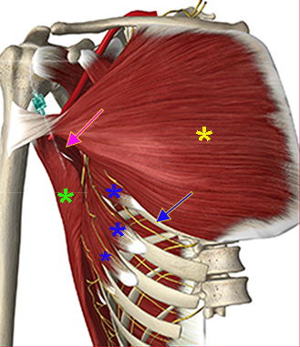
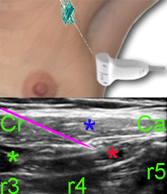
With the superficial serratus plane block, the needle tip is inserted into the fascial space between the serratus anterior muscle and the latissimus dorsi muscle caudad to the teres major muscle

Local anaesthetic injected into this fascial plane covers the thoracic intercostal nerves and their lateral cutaneous branches – and also the thoracodorsal nerve which innervates the latissimus dorsi muscle and the long thoracic nerve which innervates the serratus anterior muscle. The last mentioned nerves run alongside the thoracodorsal artery.

Blanco R, Parras T, McDonnell JG, Prats-Galino A (see ref.) have also described a serratus plane block with injection of local anaesthetic into the fascial plane deep to the serratus anterior muscle

This deeper serratus plane block also produces analgesia of the thoracic intercostal nerves and the lateral hemithorax; however, the analgesia of the deep block seems to last a shorter time than the superficial serratus plane block

Image missing
Serratus anterior m. (blue asterixs); teres major m. (magenta arrow); latissimus dorsi m. (green asterix); pectoralis major m. (yellow asterix); fifth rib (blue arrow); the needle insertion position is displayed

3 – Sonoanatomy and needle insertion

Place the patient supine

Place the probe in the parasagittal plane on the midclavicular line and the fifth rib is identified by counting from above. The rib is tracked to the midaxillary line. Rotate the probe 45 degrees from the axial plane.

The latissimus dorsi, the serratus anterior and the teres major muscles are overlying the fifth rib (see previous page). The thoracodorsal artery can be used as a marker of the fascial plane between the latissimus dorsi and serratus anterior muscles.

The needle is inserted from the anterior end of the probe and advanced in-plane

Inject 25 mL ropivacain 0.2% into the fascial plane between the serratus anterior and latissimus dorsi muscles

Image missing
The upper image shows the position of the probe and the in-plane needle orientation.
The lower image shows the fascial plane between the latissimus dorsi m. (blue asterix) and the serratus anterior m. (red asterix). Cr (cranial); Ca (caudad); r3-r5 (ribs 3-5); teres major m. (green asterix). The needle is displayed (magenta).

4 – References

Blanco R, Parras T, McDonnell JG, Prats-Galino A: Serratus plane block: a novel ultrasound-guided thoracic wall nerve block,
Anaesthesia 68: 1107-1113 (2013)

Image missing

1 – Indications

The serratus plane block produces analgesia of the lateral thoracic wall including the axilla.

The indications of the serratus plane block are postoperative analgesia after surgery of:

– lateral thoracic wall

– breast

– axilla

Image missing
The surface area of analgesia with the serratus plane block