What are B-lines?
B-lines are hyperechoic, laser-like, vertical artefacts that visually originate from the pleural line and continue to the bottom edge of the screen without fading in intensity.
B-lines and pneumothorax
Visualisation of the B-line artefact is only possible when there is no air between the two pleural blades and they are in contact with each other.
The finding of one or more B-lines can be used to rule out pneumothorax.
B-lines also play a role in diagnosing pulmonary edema (discussed later).
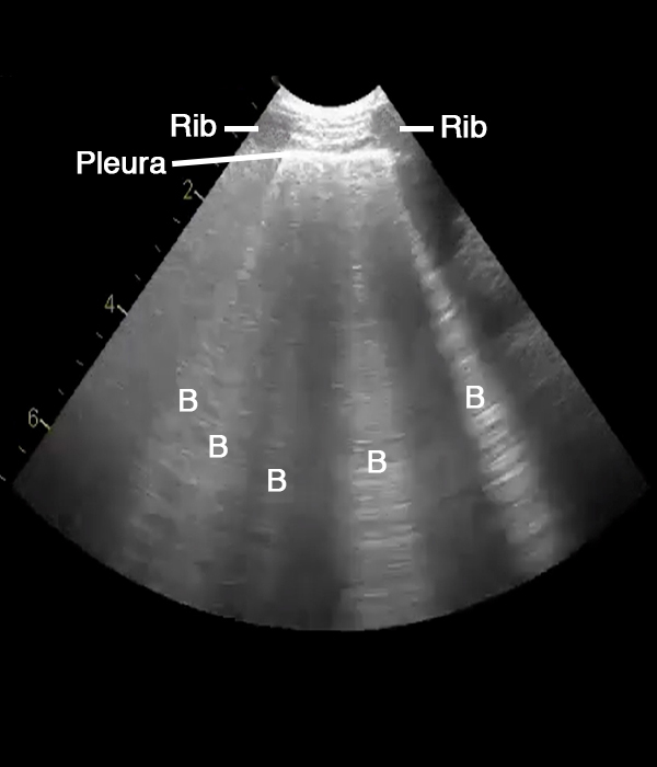
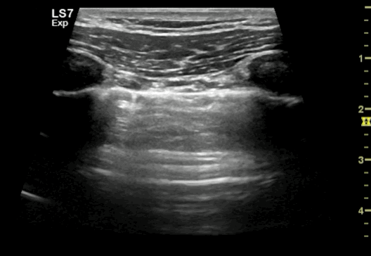
The image shows a lung ultrasound picture obtained with an abdominal curved transducer.
Rib and pleura are marked. B indicates B-lines.
Lung sliding
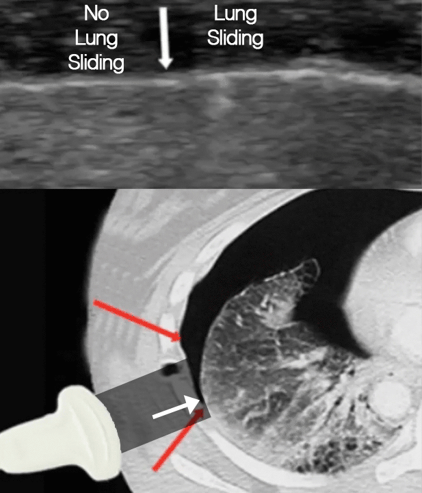
Lung sliding is a horizontal movement of the bright pleural line in synchrony with the respiratory cycle indicating sliding movement of the visceral pleura against the parietal pleura.
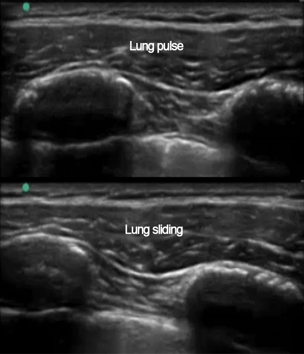
Lung pulse
Lung pulse is a variation of the same phenomenon, but is caused by projection of the heart beats through the lung tissue, causing small frequent movements of the pleural blades.
Lung pulse is only clearly seen when there is no respiration/ventilation.
Pathologies
Absence of lung sliding and lung pulse is seen in any condition causing the visceral and the parietal pleura to be either separated (e.g. pneumothorax) or joined (e.g. pleurodesis).
Presence of lung sliding or lung pulse is 100% specific and rules out pneumothorax at the location of the transducer.
Top: Lung pulse – the pulsating movement with the same frequency as the heart beating.
Bottom: Lung sliding – the sliding movement with the same frequency as the respiration.
The lung ultrasound signs Lung sliding/lung pulse, B-lines and Lung point are used in diagnosing or excluding pneumothorax. These signs are explained in the following topics.
Use the signs:
Ruling out pneumothorax:
• Presence of lung sliding/pulse or B-lines on the anterior part of the thorax
Suspicion of possible pneumothorax:
• Absence of both lung sliding/pulse and B-lines
Diagnostic of pneumothorax:
• Absence of lung sliding/pulse and B-lines on the anterior part of the thorax
• AND Presence of a “lung point”
The video shows a “lung point”, which is used for confirmation of pneumothorax. This will be explained in detail later.
Question: Can pneumothorax be confirmed or excluded using ultrasound?
Steps:
Scan the relevant part of the chest using a high-frequency linear probe.
Observe for lung signs: lung sliding/lung pulse, B-lines, and the presence of a “lung point”.
Rule out pneumothorax if lung sliding/pulse or B-lines are seen on the anterior part of the thorax.
Suspect pneumothorax if neither lung sliding/pulse nor B-lines are detected.
Confirm pneumothorax when the above signs are absent AND a lung point is present.
In-depth explanation:
Definition: Pneumothorax involves the accumulation of air in the pleural space, potentially leading to a lung collapse. Loss of contact between the visceral pleura and the parietal pleura and a gradual collapse of the affected lung can result from a continuous leak of air into the pleural space.
The application of ultrasound provides rapid pneumothorax evaluation, boasting greater sensitivity and specificity than conventional x-rays.
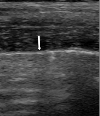
Lung point: In pneumothorax, the edge of the collapsed lung appears as a “lung point”. This point represents the transition zone between the air-filled pleura area and where the pleural blades meet. The presence of a lung point is diagnostic of a pneumothorax.
The x-ray image on the right shows pneumothorax in the right thorax The visceral pleura of the right lung is marked with white arrows
The focused question
Can pneumothorax be confirmed or excluded? The lung ultrasound signs lung sliding/lung pulse, B-lines and lung point are used in diagnosing or excluding pneumothorax. These signs are explained in the following topics.
Ruling out pneumothorax: • Presence of lung sliding/pulse or B-lines on the anterior part of the thorax
Suspicion of possible pneumothorax: • Absence of both lung sliding/pulse and B-lines
Diagnostic of pneumothorax: • Absence of lung sliding/pulse and B-lines on the anterior part of the thorax • AND Presence of a “lung point”
The sonographic lung sign
The sonographic lung sign is previously discussed in the course; refer back for detailed information.
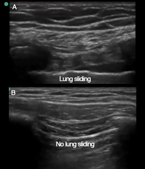
A: Normal lung sliding – PTX can be ruled out B: No lung sliding – PTX is possible
The lung point
What is a lung point? In pneumothorax, the edge of the collapsed lung will appear as a “lung point”.
The lung point represents the transition zone between the area of air-filled pleura and the reunion of the pleural blades.
The lung point moves back and forth in synchrony with the patient’s breathing.
Anterior to the lung point, with air-filled pleura, no lung sliding is seen.
Posterior to the lung point, with contacting pleural blades, normal lung sliding is seen.
Right at the lung point both can be observed as seen on the image to the right.
Lung point and pneumothorax Lung point is diagnostic of pneumothorax.
The video shows the edge of the collapsed lung during inspiration and expiration. Notice the slow movement of the lunge edge in the video, and the lack of lung sliding anterior to the lung point and the normal lung sliding posterior to the lung point. The two arrows indicate the lung edge during inspiration (top arrow) and expiration (lower arrow).The video shows a “lung point”, which is used for confirmation of pneumothorax.
Demonstrating detection of pneumothorax with ultrasound
Summary
A pneumothorax is ruled out • When lung sliding/pulse or B-lines can be seen on the anterior part of the thorax (in a patient lying on the back) A pneumothorax is suspected • When neither lung sliding/pulse nor B-lines can be seen on the anterior part of the thorax
A reliable diagnosis of pneumothorax can be established • When neither lung sliding/pulse nor B-lines can be seen on the anterior part of the thorax AND • A lung point is detected
Summary:
Key points:
A pneumothorax is ruled out when lung sliding/pulse or B-lines are evident on the anterior part of the thorax.
Suspect pneumothorax when neither lung sliding/pulse nor B-lines are detected.
Confirm diagnosis when these signs are absent AND a lung point is present.
At this juncture, you should possess a solid grasp on obtaining lung images using ultrasonography.
If you’re still unsure or lacking confidence, we recommend revisiting and reviewing the preceding topics.
However, if you’re feeling ready and confident, it’s time to advance and apply your skills in answering the focused questions.
The focused questions in PoCUS ultrasonography of the lungs are:
Can mainstem intubation be confirmed or excluded?
Can pneumothorax be confirmed or excluded?
Are signs of pulmonary edema evident or can pulmonary edema be ruled out?
Can pleural fluid be confirmed or excluded?
Are signs of atelectases present?
Helpful tips:
The answers to the first, second, and fifth questions predominantly emerge from the intercostal view.
The third and fourth questions are best addressed through the posterolateral pleural view.
At this point, you should feel confident about how to obtain the lung images.
If you do not feel confident, please review the previous topics. If you do feel confident, you can move on to answering the focused questions.
The focused questions in focused ultrasonography of the lungs are: • Can pneumothorax be confirmed or excluded? • Are signs of pulmonary edema present or can pulmonary edema be excluded? • Can pleural fluid be confirmed or excluded? • Are signs of atelectases present?
Tips: • The first two questions can be answered from the intercostal view • The latter two questions can be answered from the posterolateral pleural view
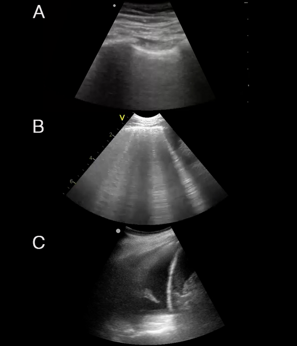
A: Anterior intercostal view. If no lung sliding is present it is suggestive of PTX B: Anterior intercostal view with b-lines, which is suggestive of pulmonary edema C: Posterolateral pleural view with pleural fluid collection
Rotation
Generally, a slight rotation of the transducer, so the OM aims more towards the mattress, can improve image quality (counter-clockwise on right side, clockwise on left side).
Fluid follows gravity
Always make sure that the transducer is placed as posterior as possible – your hand needs to touch the mattress.
That hard left side
On the left side it is generally more difficult to obtain good images than on the right side – but the tips for optimising the image are the same:
• Rotate the probe (clockwise) – maybe even more than on the right side
• Make sure the transducer is placed as posteriorly as possible.
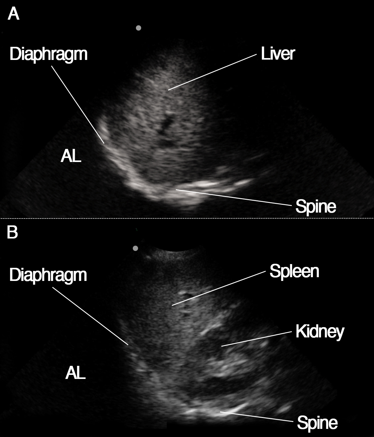
In the posterolateral pleural view, the following structures can be seen:
Right side
Liver
Kidney (not seen in the image to the right)
Diaphragm
Spine
Left side
Spleen
Kidney
Diaphragm
Spine
A: Posterolateral pleural view on the right side
B: Posterolateral pleural view on the left sideAL represents the area of the lung that is not visible because it is aerated.
The postero-lateral view is obtained by placing the transducer in the posterior axilary line, orientation marker pointing cranial. Identify liver, diaphragm and pleura and look for pleural effusion and atelectasis. The video shows you in detail how to obtain the view and identify or exclude pathology
Identifying structures
In the posterolateral pleural view, the following structures can be seen:
A: Posterolateral pleural view on the right side. B: Posterolateral pleural view on the left side. AL represents the area of the lung that is not visible because it is aerated.
Right side
Liver
Kidney (not seen in the image to the right)
Diaphragm
Spine
Left side
Spleen
Kidney
Diaphragm
Spine
Tips and tricks
Rotation
Generally, a slight rotation of the transducer, so the OM aims more towards the mattress, will assure that the transducer is aligned parallel between the ribs and avoid black shadow from underlying rib, thereby improving image quality (counter-clockwise on right side, clockwise on left side).
Fluid follows gravity
Always make sure that the transducer is placed as posterior as possible – your hand needs to touch the mattress.
That hard left side
On the left side it is generally more difficult to obtain good images than on the right side – but the tips for optimising the image are the same:
• Rotate the probe (clockwise) – maybe even more than on the right side
• Make sure the transducer is placed as posteriorly as possible.
Image shows classical intercostal view of pleura from the upper anterior part of the chest
Pleural line If the pleural line and the lung signs cannot be clearly separated from the surrounding structures: • make sure you are watching the pleural line – look into the space between two ribs, and the hyperechogenic (white) linear structure between and below the ribs is pleura – the ”BAT-sign” • Adjust the depth of the ultrasound image in order to align the pleura to the center of the ultrasound screen • try tilting the probe to obtain a 90 degree angle between the ultrasound beam and the pleural line • ask the patient to switch between breathing and holding the breath. Watch for the lung pulse and observe how lung sliding becomes more clear (breathing) when it is compared to no-sliding (breathhold) • move left /right and compare left and right side of the chest • move anterior/lateral and compare anterior with lateral part of the chest
The heart When attempting to obtain the intercostal view on the left anterior hemithorax, be aware that the heart may be in the way – in this case, move the transducer apically or laterally.
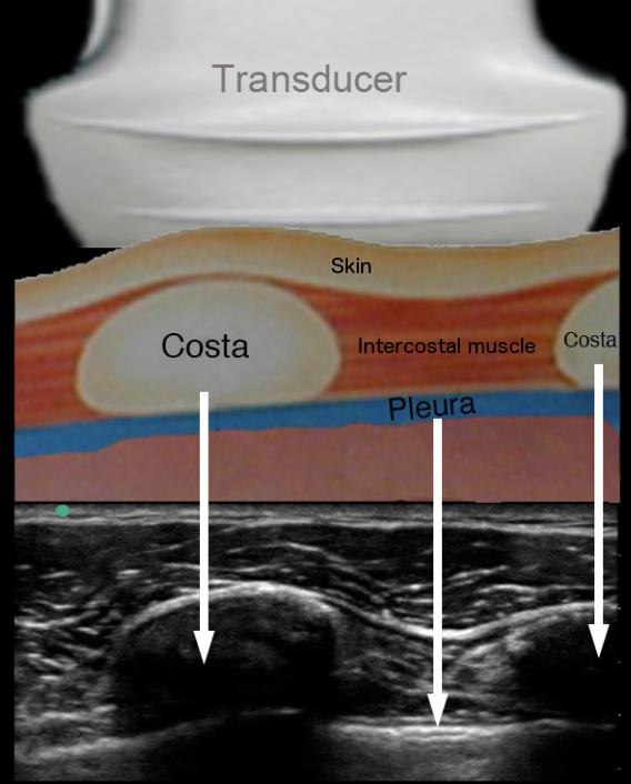
Costae
The costae are identified by the hypereechoic(white) surface and the anechoic (black) shadow below the costa.
Pleura
Pleura is seen as a hyperechogenic (white) structure between the costae.
Things are not always as they seem
OBSERVE: Image structures below the pleura line do not represent lung tissue – the air in the lungs absorbs the ultrasound waves and returns no echoes.
Structures seen in the image below the pleura line is not lung tissue but represent image artefacts from the echoes from the skin, muscle, costae and pleural line.
The image shows the presentation of the chest wall on the ultrasound screen.
The linear ultrasound probe is placed on the skin surface over the sucutaneous fat layer, costae and intercostal muscles.
The lower part of the picture shows the ultrasound image of the tissue components of the chest wall.