Lesson-Intercostal

3 – Scanning technique for ultrasound guided intercostal block

– Place the patient sitting or in the lateral decubitus position

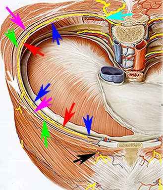
– Place the high-frequency linear probe in the parasagittal paravertebral plane approx. 10-15 cm from the sagittal midline. The lateral cutaneous branches branch off in the midaxillary line

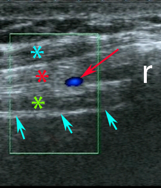
– Visualize the target intercostal space (see next page)

– Insert the needle with in-plane technique from the cranial end of the probe

– Advance the needle to the fascial plane between the internal and the innermost intercostal muscles and inject 5 mL of local anaesthetic

– The needle shold be kept meticulously in-plane in order to avoid pneumothorax

Image missing
In-plane approach to the ultrasound guided intercostal block

4 – Sonoanatomy: The intercostal nerves

The intercostal nerves are located between the innermost and the internal intercostal muscles

Typically the intercostal nerves are not visible with ultrasound. But the adjacent intercostal arteries and veins can be visualized with Color Doppler

Image missing
Intercostal artery (blue Doppler signal and red arrow), pleura (cyan arrows), innermost intercostal muscle (green asterix), internal intercostal muscle (red asterix), external intercostal muscle (cyan asterix), rib (r)

1 – Indications

– Chest drain insertion

– Rib fractures

– Rescue block with failed TAP block of the intercostal TAP plexus

Image missing
The ultrasound guided intercostal nerve block with in-plane approach

2 – Anatomy of the intercostal nerve

The spinal nerve is formed by the union of the dorsal sensory root and the ventral motor root

It passes the intervertebral foramen and branches into a ventral and a dorsal ramus. The dorsal ramus innervates the intrinsic back muscles. The ventral ramus of the intercostal nerves run anterior together with the intercostal artery and vein between two adjacent ribs sandwiched between the socalled innermost intercostal muscle (the deepest intercostal muscle layer just separated from the parietal pleura by the endothoracic fascia) and the internal intercostal muscle (the middel intercostal muscle layer). The outermost intercostal muscle layer is called the external intercostal muscle.

In the midaxillary line the intercostal nerve gives off a lateral cutaneous branch that pierces the internal and external intercostal muscles and innervates the lateral thoracic wall

Anteriorly the intercostal nerve terminates in the anterior cutaneous branch that innervates the skin covering the medial part of the trunk

Image missing
Intercostal nerve (red arrows), branching point of the dorsal and ventral rami (cyan arrow), lateral and anterior cutaneous branches (white and black arrows), innermost intercostal muscle (blue arrows), internal intercostal muscle (green arrows), external intercostal muscle (magenta arrow)