Lesson-Infraclavicular

2 – Bone distribution of the nerves from the brachial plexus

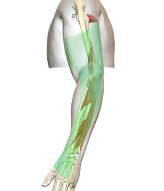
The infraclavicular brachial plexus block anaesthetize all the bones and joints of the upper limb distal to the shoulder:

– The axillary nerve innervates the head of the humerus

– The radial nerve innervates the arm, the elbow, the forearm, the hand and the
radial fingers

– The musculocutaneous nerve innervates the humerus

– The ulnar nerve innervates the elbow and the hand and the ulnar fingers

– The median nerve innervates the elbow and the forearm and the hand and the
radial fingers
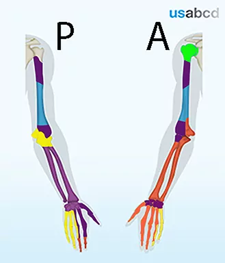
Image missing
“Bone colour” = suprascapular nerve; green = axillary nerve; purple = radial nerve;
blue = musculocutaneous nerve; yellow = ulnar nerve; orange = median nerve

3 – The muscular distribution of the nerves from the brachial plexus

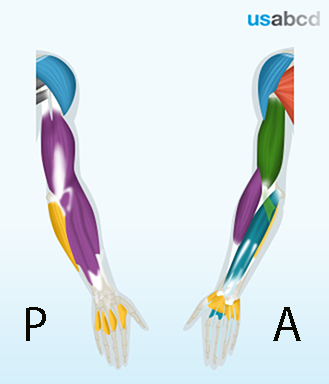
The infraclavicular brachial plexus block covers all muscles of the upper limb distal to the shoulder:

The radial nerve innervates
– the elbow extensor muscles
– the wrist extensor muscles

The musculocutaneous nerve innervates the elbow flexor muscles

The ulnar nerve innervates the intrinsic muscles of the hand including the medial finger flexors

The median nerve innervates the wrist flexor muscles including the lateral finger flexors

Image missing
Blue: axillary nerve (not covered by an infraclavicular block); green: musculocutaneous nerve; purple: radial nerve; blue: median nerve; yellow: ulnar nerve; gray: teres minor and major nerves (axillary and subscapular nerves – not covered by an infraclavicular block); orange: pectoral nerves.

4 – The cutaneous distribution of the nerves from the brachial plexus

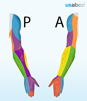
The infraclavicular brachial plexus block provides cutaneous anaesthesia of the entire upper limb

The radial nerve innervates the skin of the back of the brachium and the lateral hand

The musculocutaneous nerve innervates the skin of the lateral antebrachium

The ulnar nerve innervates the skin of the medial hand and fingers

The median nerve innervates the skin of the anterior part of the lateral hand and fingers and the distal dorsal part of the second and third finger

The skin of the medial arm and forearm is innervated by the medial cutaneous nerves of the arm and forearm. They are both derived from the medial cord of the brachial plexus and covered by the infraclavicular brachial plexus block

Image missing
Blue (shoulder): axillary nerve; purple: radial nerve; orange (arm): medial cutaneous nerve of the arm; yellow: musculocutaneous nerve; green: medial cutaneous nerve of the forearm; blue (hand): ulnar nerve; orange (hand): median nerve

1 – The infraclavicular block of the brachial plexus

The infraclavicular block anaesthetises the brachial plexus at the level of the cords

The upper limb is innervated by nerves from the brachial plexus (BP). The BP is derived from five spinal nerve roots C5-T1

The superior trunk (ST) comes from C5 and C6. The middle trunk comes from C7 (MT). The inferior trunk (IT) comes from C8 and T1. The anterior branches from ST and MT unite to become the lateral cord. The anterior branch of IT is the medial cord. The three posterior branches of ST, MT and IT unite to become the posterior cord

The axillary nerve and the radial nerve come from the posterior cord. The musculocutaneous nerve and part of the median nerve come from the lateral cord. The ulnar nerve and part of the median nerve come from the medial cord. The medial cutaneous nerves of the arm and the forearm branches off the medial cord. The suprascapular and axillary nerves are not anaesthetised by the infraclavicular block

Image missing
ST/MT/IT: superior/middle/inferior trunks; NERVES: axillary (AX), radial (R), musculocutaneous nerve; (MC), median (M); ulnar (U); medial cutaneous nerves of the arm and forearm (CMAB); suprascapular (SS); red/yellow asterixs: anterior/posterior divisions; PC/LC/MC: posterior/lateral/medial cords

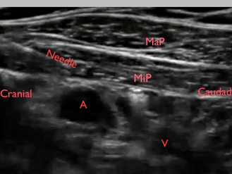
15 – Injection of local anaesthetic around the medial cord

The medial cord is located between the axillary artery and vein around the two o’clock position of the artery

The local anaesthetic does not always spread around the artery to the two o’clock position from a seven o’clock point of injection

In that case the needle tip should be repositioned in front of the axillary artery to the interspace between the axillary artery and vein and local anaesthetic should be injected periarterially

Click on the VIDEO CLIP button to view the video

Image missing
Periarterial injection of local anaesthetic between the axillary artery and vein in order to cover the medial cord of the brachial plexus
MaP = major pectoral muscle; MiP = minor pectoral muscle; A = axillary artery; V = axillary vein

16 – Colour Doppler

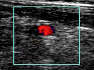
Colour Doppler can be used to visualise the proxy marker – the axillary artery – and blood vessels that you do not want to perforate with the needle

It is also possible to use colour Doppler to visualise the injection of local anaesthetic

In that case it is an advantage to lower the PRF (Pulse Repetition Frequency) in order to enhance the visibility of the injectate

Click on the VIDEO CLIP button to view the video

Image missing
The video shows the pulsating colour Doppler signal from the axillary artery

17 – Avoid injecting air perineurally

Before inserting the needle through the skin, the hose and the needle should be filled with saline or local anaesthetic

Just after insertion of the needle 1/2 ml of fluid should be flushed through the hose and needle to make sure that no air is contained in the “system”

Click on the VIDEO CLIP button to view the video

Image missing
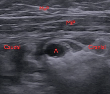
The video shows the blurring effect of injected air during the performance of an infraclavicular nerve block
A = axillary artery; MaP = major pectoral muscle; MiP = minor pectoral muscle

14 – The infraclavicular block with repositioning of the needle tip

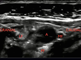
Repositioning of the needle tip is oftentimes necessary in order to cover all three cords around the axillary artery from two o’clock to ten o’clock

Care should be taken not to pierce the axillary vein

Click on the VIDEO CLIP button to view the video

In the video: Notice that local anaesthetic is injected on the wrong side of the clavipectoral fascia which has to be penetrated by the needle before local anaesthetic spreads appropriately around the medial cord

Image missing
The video shows repositioning of the needle tip during an infraclavicular block
First, local anaesthetic is injected around the lateral cord in the ten o’clock position
Then, the needle is relocated in front of the axillary artery in order to reach the medial cord