Dolan J, Williams A, Murney E, Smith M, Kenny GNC: Ultrasound guided fascia iliaca block: A comparison with the loss of resistance technique. Reg Anesth Pain Med 33: 526-531 (2008)
Dolan J, Williams A, Murney E, Smith M, Kenny GNC: Ultrasound guided fascia iliaca block: A comparison with the loss of resistance technique. Reg Anesth Pain Med 33: 526-531 (2008)
Postoperative analgesia after:
– hip surgery
– anterior thigh surgery
– knee surgery
Only when a femoral nerve block and/or a lateral femoral cutaneous nerve block is not feasible
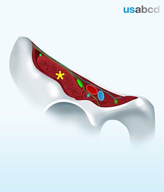
The fascia iliaca is the muscle fascia of the iliacus muscle and is continuous with the fascia of the psoas major muscle
Laterally it is attached to the iliac crest
The femoral as well as the lateral femoral cutaneous nerves are located underneath the fascia iliaca
The idea of the fascia iliaca block is to inject a large volume of local anaesthetic midways between the femoral and lateral femoral cutaneous nerves in order to obtain lateral and medial spread that will cover both nerves
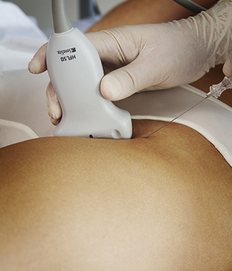
– Place the patient supine
– Place the high-frequency linear probe in the femoral crease, lateral to the femoral artery
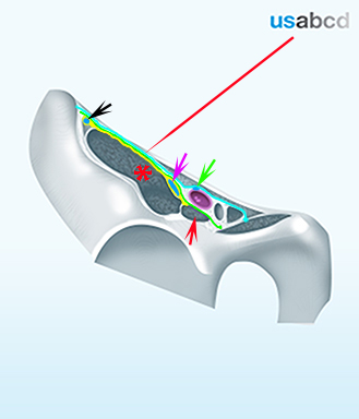
– Identify the femoral artery, the iliopsoas muscle, the fascia iliaca, and the sartorius muscle by sliding lateral (see next page)
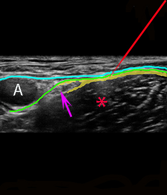
– Insert the needle from the lateral end of the probe with in-plane technique and pierce the fascia iliaca (usually with a palpable “pop”
– Inject local anaesthetic with visible spread between the muscle and the fascia iliaca without resistance and no pain

The needle is advanced through the fascia lata and the iliaca fascia – but not into the muscle
Local anaesthetic is injected into the fascial plane between the iliaca fascia and the muscle