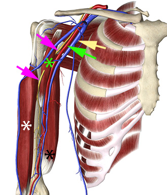
The musculocutaneous (MC) nerve origins from the lateral cord of the brachial plexus. Proximal to the lower border of the axilla (= the lower margin of the teres major muscle) the trajectory of the MC nerve deviates from the axillary artery and veins and the nerve exits the axilla by piercing the coracobrachialis muscle and descends and ramifies in the fascial plane between the coracobrachialis muscle and the short head of the biceps brachii muscle
This means that the MC nerve has to be blocked separately in the fascial plane between the coracobrachialis muscle and the short head of the biceps brachii muscle with the axillary block
The short head of the biceps muscle has been removed exposing the long head of the biceps (white asterix) and the piercing of the coracobrachialis muscle (green asterix) by the musculocutaneous nerve (magenta arrows)
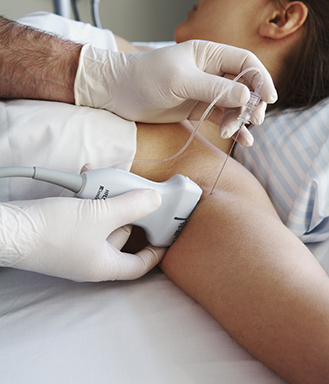
– Place the patient supine with the arm abducted and elbow bend 90 degrees
– Place a high-frequency linear probe axially in the axillary crease to obtain a cross-sectional view of the axillary artery
– Optimize depth, gain and focus
– Insert the needle with in-plane technique from the cranial end of the probe
– Block the nerves individually with 3-5 mL of local anaesthetic per nerve or deposit local anaesthetic periarterially; always block the musculocutaneous nerve separately
Place the patient supine with abducted arm and bend elbow and insert the needle with in-plane approach
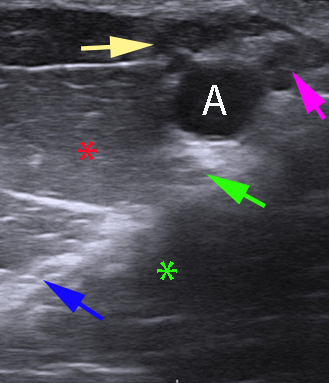
Visualise the median, ulnar, radial, and musculocutaneous nerves around the pulsatile axillary artery and the veins
If the nerves are invisible they can be traced from the elbow or local anaesthetic can be deposited periarterially. If a nerve (typically the radial nerve) cannot be visualized, it can be rescue blocked proximal to the elbow.
The median nerve is typically located lateral and superficial in relation to the axillary artery, the ulnar nerve is medial and the radial is deep to the artery. These three nerves typically are sonographically heterogenous. The musculocutaneous nerve is apart from the artery – typically in the fascial plane between the short head of the biceps muscle and the coracobrachialis muscle.
The veins are compressed and become invible when the probe pressure is increased; the blood vessels can be visualised with colour Doppler
Axillary artery (A), musculocutaneous nerve (blue arrow), radial nerve (green arrow), median nerve (yellow arrow), ulnar nerve (magenta arrow), short head of the biceps brachii muscle (red asterix), corabrachialis muscle (green asterix).
The axilla extends from the first rib to the lower margin of the posterior axillary fold (= the lower border of the teres major muscle)
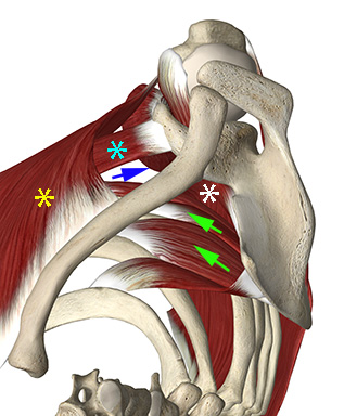
Posterior wall is the subscapularis, teres major and latissimus dorsi muscles
Anterior wall is the major and minor pectoral muscles
The lateral wall is the humerus and the tendons of the short head of the biceps muscle and the coracobrachialis muscle
Posterior wall (white asterix): subscapularis, teres major and latissimus dorsi muscles. Anterior wall: major & minor pectoral muscles (yellow and cyan asterixs). Lateral wall: humerus and the tendons of the short head of the biceps and coracobrachialis muscles (blue arrow). Medial wall: serratus anterior (green arrows).
The axilla (armpit) is bound medially by the rib cage and the serratus anterior muscles, anteriorly by the anterior axillary fold (the major and minor pectoral muscles – yellow and cyan asterixs in the picture), laterally by humerus and the tendons of the coracobrachialis muscle (green asterix) and of the short head of biceps brachii muscle (blue asterix), and posteriorly by the subscapularis (white asterix) and the posterior axillary fold
The posterior axillary fold is the latissimus dorsi muscle (not shown) and the teres major muscle (magenta asterix)
The triceps muscle is shown with a black asterix
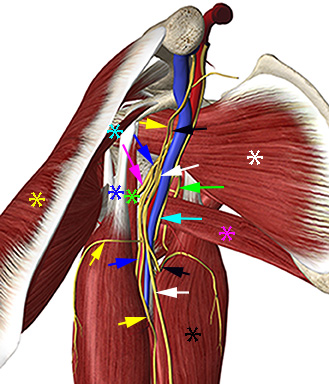
The ultrasound-guided axillary block is performed in cross-section along the lower border of the teres minor muscle, where the ulnar nerve, median nerve, radial nerve, and medial and antebrachial cutaneous nerves run in close proximity to the axillary artery and veins (see figure and text). The musculocutaneous nerve has branched off and has to be blocked separately at this level. The axillary nerve is not blocked with the axillary block.
The axilla from a medial view. Musculocutaneous nerve (magenta arrow), median nerve (blue arrow), ulnar nerve (white arrow), radial nerve (cyan arrow), axillary nerve (green arrow), medial brachial and antebrachial cutaneous nerves (black and yellow arrows)