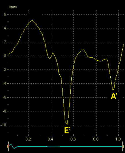
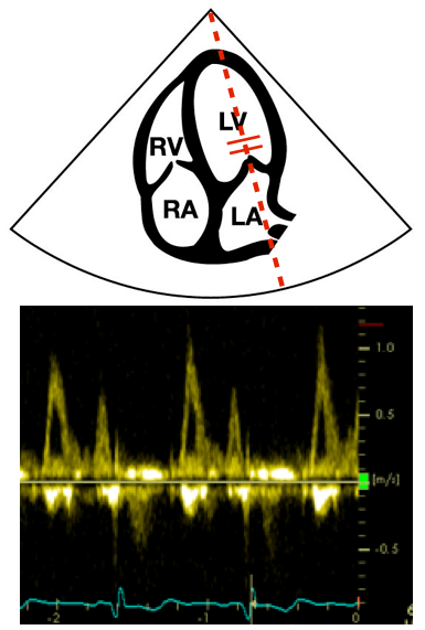
Measuring the myocardial velocities in the mitral annulus by means of tissue Doppler imaging (TDI) adds significant value to the evaluation of the left ventricular diastolic function
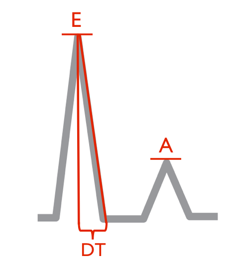
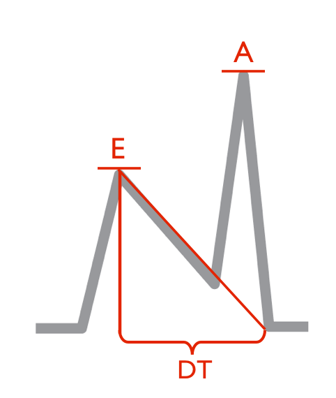
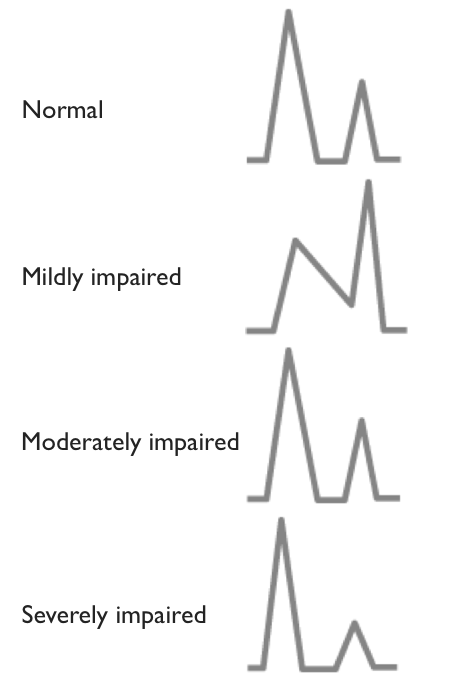
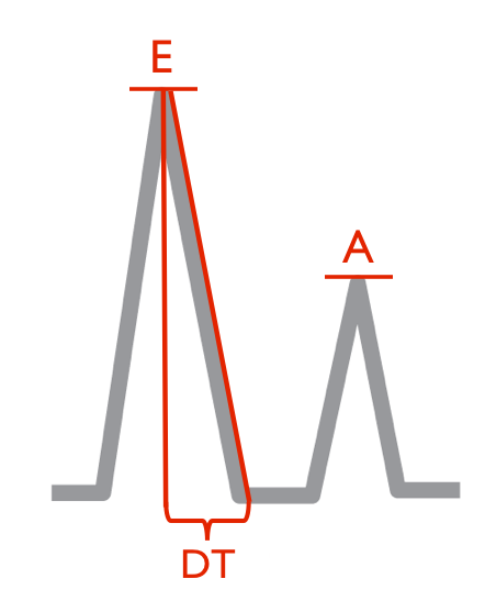
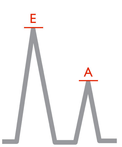
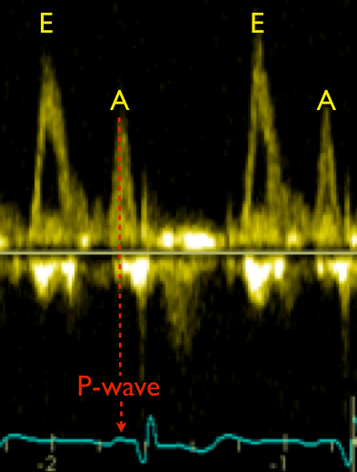
Tissue Doppler recording is characterised by two diastolic peak velocities, the E’ (E prime) as an early event and the A’ (A prime) during atrial contraction
Normally E’ > A’ with a ratio of approximately 1-2
Like for the transmitral E and A, the ratio reverses progressively as diastolic dysfunction develops