FATE

Manipulating the probe


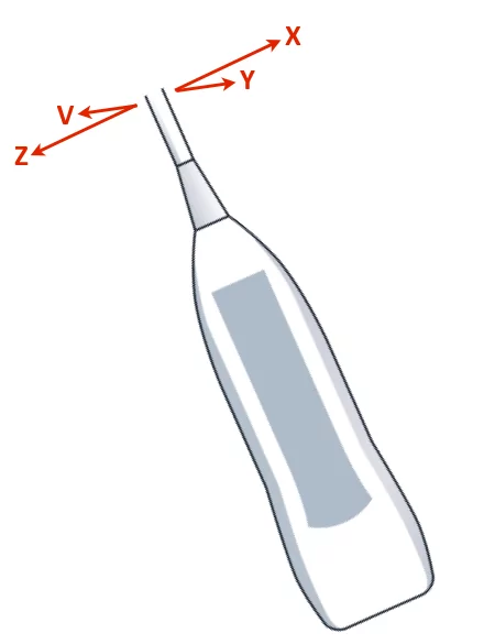
A terminology to describe the movement of the probe during scanning is important in order to optimise the image

During the workshop it makes it easier for the supervisor to give instructions without touching the probe which is crucial for the novice in order to achieve practical skill

There are many available descriptions of the method of manipulation of the probe
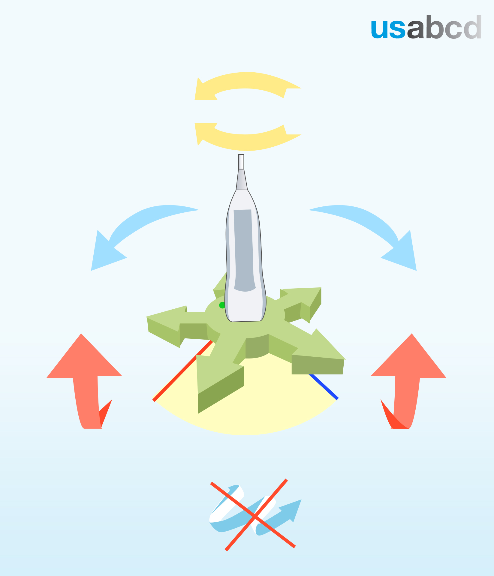
We recommend using:
1. rotate
2. tilt
3. slide

These movements are applicable to all scanning locations

As a novice – always move the probe in only one direction at a time

Image missing
Movements in three different spatial directions.The icon at the bottom indicates that movement should only take place in one plane at the time.

Instruction in manipulation of the probe


During the workshops the supervisors are encouraged to give instructions with reference to the tail of the transducer and a secondary reference point when appropriate

Examples of secondary reference points:
1. Anatomical landmarks in relation to the volunteer or patient
2. Physical landmarks like the bed
3. Spatial orientation: left/right – upwards/downwards – medial/lateral

When complying with an instruction, never do any movements in other directions

Image missing
Transducer manipulations with reference to the tail of the probe and four different secondary reference points: X, Y, Z ,V

Focus Assessed Transthoracic Echocardiography (FATE)


FATE is an acronym for “Focus Assessed Transthoracic Echocardiography”

FATE is the original focused echo protocol for all physicians – practiced since 1989

FATE is easily and quickly learned and can be applied in all clinical scenarios – e.g. pre-, intra-, and postoperatively, intensive care medicine, emergencies, and resuscitation

FATE can be performed with the patient in the sitting position

The FATE card (see figure) also encompasses quick guidance in the interpretation of the echocardiographic findings and their application in the clinical context

FATE can be performed in all locations: OR, ICU, emergency room, pre-hospital, ambulance, in-flight, patient’s home – there are no limitations

Image missing
You can download the FATE card from: usabcd.org/FATE-card

FATE levels of training


There are currently two available levels of FATE training

Basic FATE (Level 1)
Based on two-dimensional imaging of the four easiest cardiac views, IVC and pleural supplemented by M-mode, the subject learns cardiac function and the most important pathology seen in critical care

No precourse qualifications are required

Advanced FATE (Level 2)
Basic FATE supplemented with extended cardiac views and Doppler ultrasound for cardiac output and pressure measurements, together with assessment of LV diastolic function. This module requires completion of Basic FATE level of training or a similar level

Image missing
Requirements for Level 1 certification as suggested by USabcd

The FATE card


The course is based on the famous FATE card, either the laminated hard copy or apps for iPhone or Android

You should keep the FATE card in front of you during the entire course

You can download the FATE card here

The FATE Card app is also available in the Appstore and Google Play

Image missing

Course objective for Basic FATE


The objective for the Basic FATE course is to provide the attendees with basic echocardiographic skills for the non-cardiologist in order to perform cardiopulmonary evaluation and optimization

After the course:

• You will know the normal sonographic features of the heart, inferior vena cava and pleura
• You will be able to obtain the basic FATE views
• You will be familiar with M-mode (motion-mode) for assessment of
cardiac dimensions and function
• You will know about and can recognize the characteristics of the most
important pathologies
• You can put the echocardiographic findings into the clinical context

Image missing
The image shows the four images obtained from the three cardiac windows in the FATE examination.

Equipment and skills


Basic understanding of the ultrasound system user controls is a prerequisite for learning FATE

Before you start clinical FATE scanning it is important to become familiar with the probe (or transducer) and the user interface of the ultrasound system

Image missing