FM-quiz FATEadv
FM-quiz FATEadv
Quiz Summary:
0 of 52 questions completed
Questions:
Information
You have already completed the quiz before. Hence you can not start it again.
Quiz is loading…
You must sign in or sign up to start the quiz.
You must first complete the following:
Results
Results
0 of 52
Time has elapsed
0 of 0 point(s), (0)
0 of 0, (0)
Essay(s) Pending: 0 (Possible Point(s): 0)
Categories
- FillInTheBlank 0%
- Multiple Choice 0%
- Summary 0%
- 1
- 2
- 3
- 4
- 5
- 6
- 7
- 8
- 9
- 10
- 11
- 12
- 13
- 14
- 15
- 16
- 17
- 18
- 19
- 20
- 21
- 22
- 23
- 24
- 25
- 26
- 27
- 28
- 29
- 30
- 31
- 32
- 33
- 34
- 35
- 36
- 37
- 38
- 39
- 40
- 41
- 42
- 43
- 44
- 45
- 46
- 47
- 48
- 49
- 50
- 51
- 52
- Current
- Review
- Answered
- Correct
- Incorrect
-
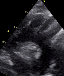
1 of 52Question1PSAX view at the mitral level. Which of the following statements about the PSAX view at the mitral level are correct?
 CorrectIncorrect
 -
2 of 52Question2TEST 1: Assessment of cardiac output using Doppler. Which statements are true regarding the assessment of cardiac output using Doppler in the left ventricle outflow tract?
 CorrectIncorrect
 -
3 of 52Question3PSAX view at the aorta level. Which of the following statements about the PSAX view at the aortic level are correct?
 CorrectIncorrect
 -
4 of 52Question4TEST 2: Assessment of cardiac output using Doppler. Which of the following statements regarding the assessment of volume flow with Doppler are correct?

 CorrectIncorrect
 -
5 of 52Question5Key points of the suprasternal view. Which of the following statements about suprasternal view are correct? Select all that apply.
 CorrectIncorrect
 -
6 of 52Question6TEST 3: Cardiac output – CSA assessment. Where should the diameter be measured for the assessment of CO in the LVOT?
 CorrectIncorrect
 -
7 of 52Question7Cardiac output. Which of the following statements about cardiac output are correct? Select all that apply.
 CorrectIncorrect
 -
8 of 52Question8Doppler modalities 1. Which of the following statements about the different Doppler modalities are correct?
 CorrectIncorrect
 -
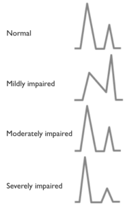
9 of 52Question9Diastolic function – mitral inflow curve. Which of the following statements about the assessment of diastolic function are correct? Select all that apply.
 CorrectIncorrect
 -
10 of 52Question10Doppler modalities. Which of the following statements about the different Doppler modalities are correct? Select all that apply.
 CorrectIncorrect
 -
11 of 52Question11Diastolic function – mitral inflow curve. Which of the following statements regarding the assessment of diastolic function are correct? Select all that apply.
 CorrectIncorrect
 -
12 of 52Question12Pressure estimation. Which of the following statements about pressure estimation are correct? Select all that apply.
 CorrectIncorrect
 -
13 of 52Question13Diastolic function. Which of the following statements about diastolic function are correct? Select all that apply.
 CorrectIncorrect
 -
14 of 52Question14Pressure estimation. Which of the following statements about Doppler pressure estimation are correct? Select all that apply.

 CorrectIncorrect
 -
15 of 52Question15Diastolic function – PWD and TDI. Which of the following statements regarding diastolic function assessed by pulsed wave and tissue Doppler are correct?
 CorrectIncorrect
 -
16 of 52Question16Pressure estimation. Which of the following statements about the pressure estimation are correct? Select all that apply.
 CorrectIncorrect
 -
17 of 52Question17Diastolic function – PWD and TDI. Which of the following statements regarding diastolic function assessed by pulsed wave and tissue Doppler are correct?
 CorrectIncorrect
 -
18 of 52Question18Colour gain. The picture shows two ultrasound images, where B is an optimised image of A. How is B optimised? Only one true answer
 CorrectIncorrect
 -
19 of 52Question19TEST 4: Spectral Doppler curves obtained across the aortic valve. The image shows 3 different spectral Doppler curves obtained across the aortic valve. Which of the following statements are correct?
 CorrectIncorrect
 -
20 of 52Question20Doppler modalities. Which of the statements about Doppler modalities are correct? Select all that apply.
 CorrectIncorrect
 -
21 of 52Question21Aortic peak pressure. Which of the following statements about the aortic peak pressure are correct? Select all that apply.
 CorrectIncorrect
 -
22 of 52Question22Right place for measuring the diameter of IVC. Where should the IVC diameter be measured?
 CorrectIncorrect
 -
23 of 52Question23Spectral Doppler curves obtained across the aortic valve. The image shows 3 different spectral Doppler curves obtained across the aortic valve. Which of the following statements are correct? Select all that apply.
 CorrectIncorrect
 -
24 of 52Question24IVC statements 1. Which of the following statements about the IVC long axis view are correct ?
 CorrectIncorrect
 -
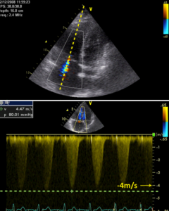
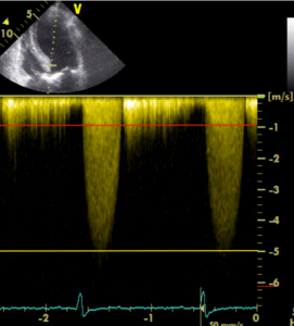
25 of 52Question25TEST 7: CW Spectral Doppler curves obtained across the aortic valve. The image shows a CW spectral Doppler curve across the aortic valve. Which of the following statements are correct?
 CorrectIncorrect
 -
26 of 52Question26
IVC statements 2
Which of the following statements about the IVC are correct? Select all that apply.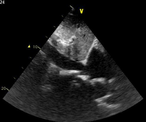
 CorrectIncorrect
 -
27 of 52Question27CW Spectral Doppler curves obtained across the tricuspid valve. The image shows a CW spectral Doppler curve across the tricuspid valve. Which of the following statements are correct? Select all that apply.
 CorrectIncorrect
 -
28 of 52Question28Key points of the extended apical views. Which of the following statements about the apical 2 chamber and long axis views are correct? Select all that apply.
 CorrectIncorrect
 -
29 of 52Question29Right side peak pressure estimation. Which statements are correct about right side peak pressure from this 4 ch view? Select all that apply.
 CorrectIncorrect
 -
30 of 52Question30Key points of the extended apical views 2. Which of the following statements about the apical 5 chamber view are correct?
 CorrectIncorrect
 -
31 of 52Question31TEST 10: CW Doppler curves across the tricuspid valve. A-C represents CW Doppler curves across the tricuspid valve, which of the following statements are correct ?
 CorrectIncorrect
 -
32 of 52Question32
2D sonoanatomy of IVC and surrounding structures
 Test yourself: Insert missing letters. In the image each letter labels a structure. Please compare the image with the statements. Insert the correct letter missing in each statement and click the
-
Shows Deep part of the liverShows IVC – Right atrial junctionShows Lung tissueShows Right atriumShows Superficial part of the liverShows Inferior vena cava (IVC)
Correct 5 / 5 PointsIncorrect / 5 Points33 of 52Question33Sonoanatomy 1
 Test yourself: Insert missing letters. In the image each letter labels a structure. Please compare the image with the statements. Insert the correct letter missing in each statement and click the
-
Shows Left atriumShows Inferior-medial wallShows Left ventricleShows Anterior wall of the left ventricleShows Coronary sinusShows Apex
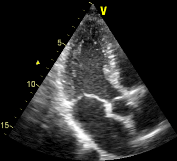
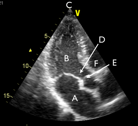
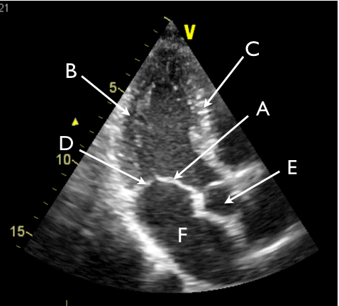
Correct 5 / 5 PointsIncorrect / 5 Points34 of 52Question34Sonoanatomy 2
 Test yourself: Insert missing letters. In the image each letter labels a structure. Please compare the image with the statements. Insert the correct letter missing in each statement and click the
-
Shows Anterior mitral leafletShows Posterior mitral leafletShows Anterior wall of the left ventricleShows Inferior-medial wallShows ApexShows Left atrium
Correct 5 / 5 PointsIncorrect / 5 Points35 of 52Question35Sonoanatomy 3
 Test yourself: Insert missing letters. In the image each letter labels a structure. Please compare the image with the statements. Insert the correct letter missing in each statement and click the
-
Shows Left atriumShows Right ventricleShows Left ventricular outflow tractShows Left ventricleShows ApexShows Aortic valve
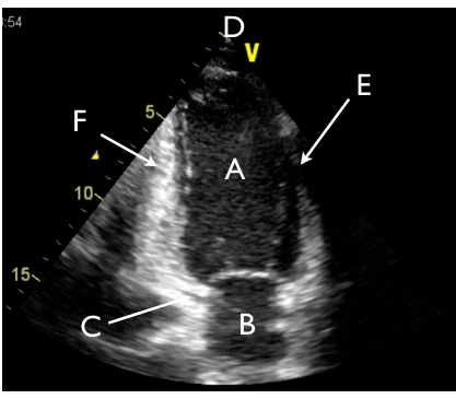
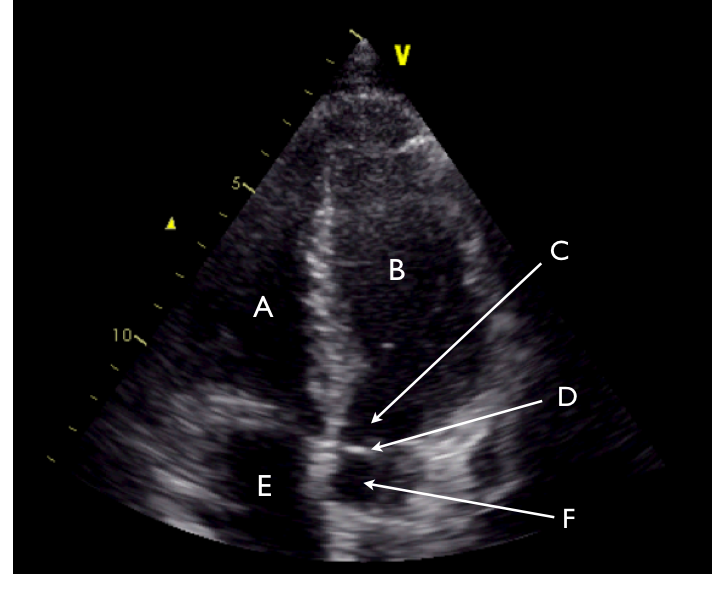
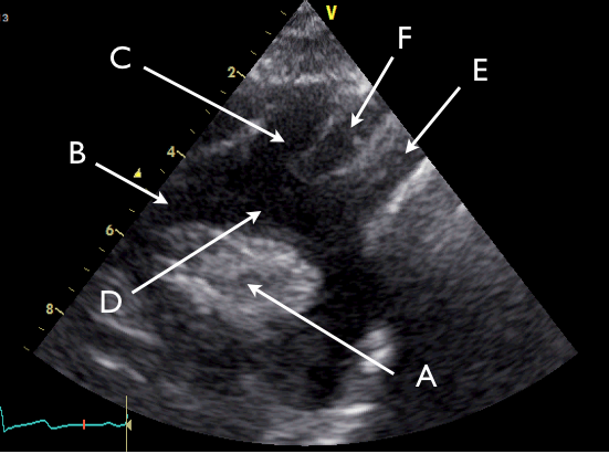
Correct 5 / 5 PointsIncorrect / 5 Points36 of 52Question36Sonoanatomy 4
 Test yourself: Insert missing letters. In the image each letter labels a structure. Please compare the image with the statements. Insert the correct letter missing in each statement and click the
-
Shows LV Inferior lateral wallShows Left atriumShows Ascending aortaShows Posterior mitral leafletShows Anterior-medial wallShows Anterior mitral leaflet
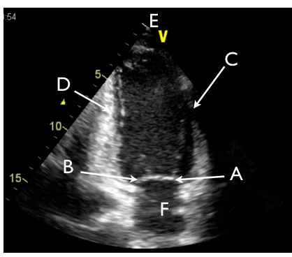
Correct 5 / 5 PointsIncorrect / 5 Points37 of 52Question37Sonoanatomy 5
 Test yourself: Insert missing letters. In the image each letter labels a structure. Please compare the image with the statements. Insert the correct letter missing in each statement and click the
-
Shows Right atriumShows Right ventricleShows Left ventricleShows Ascending aortaShows Left ventricular outflow tractShows Aortic valve
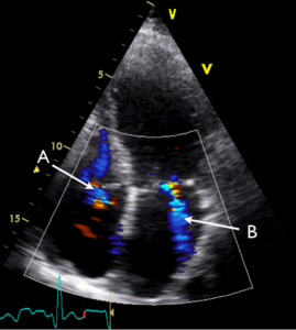
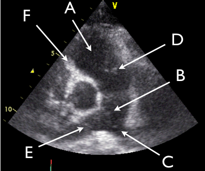
Correct 5 / 5 PointsIncorrect / 5 Points38 of 52Question38PSAX view mitral level test: 2D sonography of the cardiac structures – 1
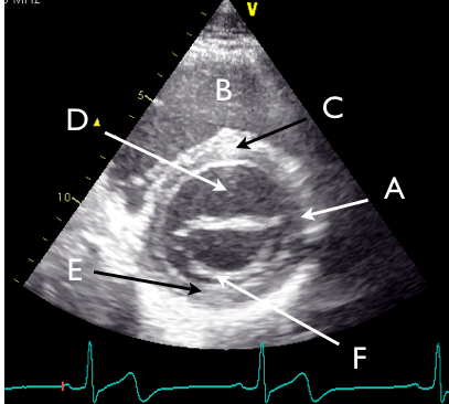 Test yourself: Insert missing letters. In the image each letter labels a structure. Please compare the image with the statements. Insert the correct letter missing in each statement and click the
-
Shows Anterior mitral leafletShows Posterior wallShows Right ventricleShows Interventricular septumShows Lateral wallShows Posterior mitral leaflet
Correct 5 / 5 PointsIncorrect / 5 Points39 of 52Question39PSAX view mitral level test: 2D sonography of the cardiac structures – 2
In the image each letter labels a structure. Insert the correct letters for each statement and click “Next”.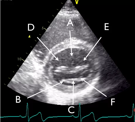
-
shows A3shows A2shows P3shows A1shows P2shows P1
CorrectIncorrect40 of 52Question40PSAX pulmonary artery: 2D sonography of the cardiac structures
 Test yourself: Insert missing letters. In the image each letter labels a structure. Please compare the image with the statements. Insert the correct letter missing in each statement and click the
-
Shows Tricuspid valveShows Right pulmonary arteryShows Pulmonary valveShows Left pulmonary arteryShows Right ventricleShows Pulmonary trunk
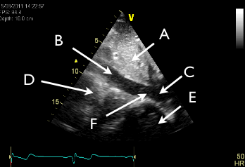
Correct 5 / 5 PointsIncorrect / 5 Points41 of 52Question41PSAX aortic level: 2D sonography of the cardiac structures
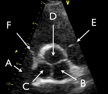 Test yourself: Insert missing letters. In the image each letter labels a structure. Please compare the image with the statements. Insert the correct letter missing in each statement and click the
-
Shows Right atriumShows Tricuspid valveShows Right aortic cuspShows Pulmonary valveShows Left aortic cuspShows Non-coronary cusp
Correct 5 / 5 PointsIncorrect / 5 Points42 of 52Question42Suprasternal view: 2D sonography of the cardiac structures
 Test yourself: Insert missing letters. In the image each letter labels a structure. Please compare the image with the statements. Insert the correct letter missing in each statement and click the
-
Shows Left common carotid arteryShows Ascending aortaShows Left subclavian arteryShows Aortic archShows Brachiocephalic trunkShows Right pulmonary aretry
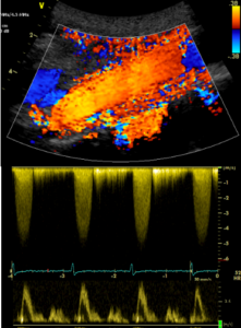
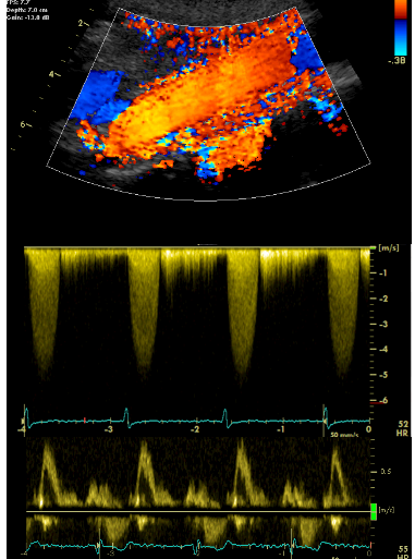
Correct 5 / 5 PointsIncorrect / 5 Points43 of 52Question43Understanding the Doppler image
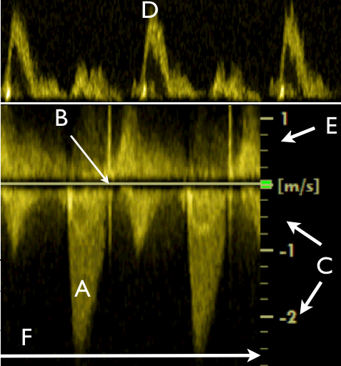 Test yourself: Insert missing letters. In the image each letter labels a structure. Please compare the image with the statements. Insert the correct letter missing in each statement and click the
-
Shows Continuous wave spectral Doppler curvesShows Baseline = 0 velocity lineShows Time lineShows Pulsed wave spectral Doppler curvesShows Scale of negative velocitiesShows Scale of positive velocities
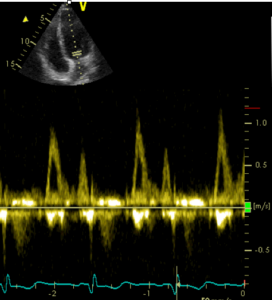
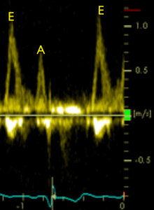
Correct 5 / 5 PointsIncorrect / 5 Points44 of 52Question44Diastolic Function – PWD and TDI
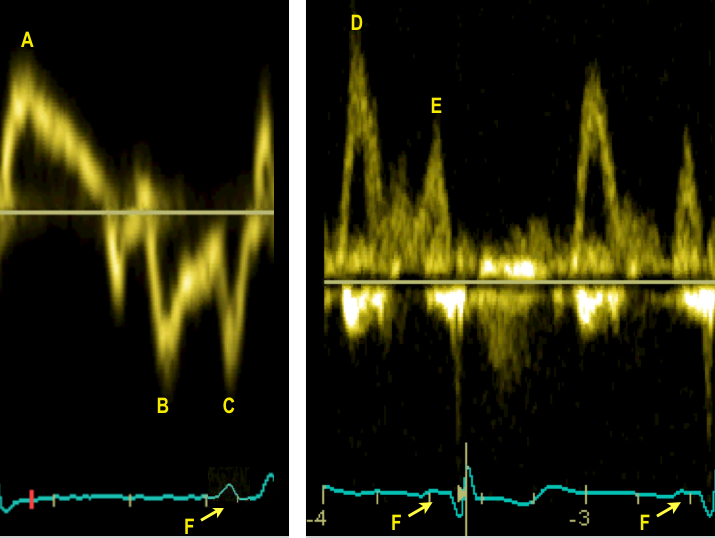 Test yourself: Insert missing letters. In the image each letter labels a structure. Please compare the image with the statements. Insert the correct letter missing in each statement and click the
-
Shows A’ = diastolic atrial contraction (tissue Doppler at mitral annulus)Shows E’ = diastolic early velocity (tissue Doppler at mitral annulus)Shows A = atrial contraction (mitral inflow with pulsed wave Doppler)Shows E = early diastolic velocity (mitral inflow with pulsed wave Doppler)Shows S = systolic peak velocity (tissue Doppler at mitral annulus)Shows P-wave in the ECG (synchronized with A and A’)
Correct 5 / 5 PointsIncorrect / 5 Points45 of 52Question45IVC summary questions
Match the phrase at the top with the correct option below to form true statements by dragging the answer into the answer box. Click “Next” when finished.Sort elements
- will display the aorta inferior to the IVC on the screen (if visible)
- will display the aorta on the screen when the imaging plane is directed to the patient’s left
- in healthy spontaneously breathing subjects
- in patients with pulmonary embolus, right heart failure or pulmonary hypertension
- can be assessed on both M-mode and 2D images
- can be made offline on a dynamic 2D image on some machines
-
Imaging the IVC through the liver from position 4
-
A correct image of the IVC from position 1
-
The IVC generally changes with respiration
-
The IVC is often dilated and changes minimally with respiration
-
Dynamic changes of the IVC with respiration
-
Anatomical M-mode
CorrectIncorrect46 of 52Question46Match the phrases on the left side with the phrases on the right side to form true statements. Click “Submit” when finishedhttps://usabcd.org/wp-content/uploads/FM_media/FATEadv/FATEadv_Q_M4_P44?
Sort elements
- the transducer should be rotated 60 degrees clockwise
- the transducer should be tilted without any rotation (tail of the transducer down)
- the transducer should be kept constant apart from a counterclockwise rotation
- is not suitable for the evaluation of the ascending aorta
- is suitable for the evaluation of left ventricle outflow tract and ascending aorta
-
To obtain the apical 2Ch view when starting with the apical long axis view
-
To obtain the apical 5Ch view from the apical 4Ch view
-
To obtain the apical 2Ch and long axis view when starting from the apical 4Ch view
-
The apical 2Ch view
-
The apical long axis view
CorrectIncorrect47 of 52Question47Match the phrases on the left side with the phrases on the right side to form true statements. Click “Submit” when finishedhttps://usabcd.org/wp-content/uploads/FM_media/FATEadv/FATEadv_Q_M5_P27?
Sort elements
- all three aortic cusps are generally visible
- both the anterior and posterior mitral leaflet are generally visible
- the right ventricle is generally visible as a common feature
- appear in the far field and on the right side of the screen
- appear in the near field
-
In the parasternal SAX view at aortic level
-
In the parasternal SAX view at mitral level
-
In the parasternal SAX view at the aortic and mitral levels
-
In the parasternal SAX view at aorta level the pulmonary artery bifurcation
-
In the parasternal SAX view at mitral level the right ventricle
CorrectIncorrect48 of 52Question48Match the phrases on the left side with the phrases on the right side to form true statements. Click “Submit” when finishedhttps://usabcd.org/wp-content/uploads/FM_media/FATEadv/FATEadv_Q_M6_P11?
Sort elements
- the ascending aorta, aortic arch and the descending aorta are seen in long axis
- aortic pressure drop can be estimated
- is generally visible under the aortic arch
- is generally not visible
- most likely appear on the left side of the screen in the correctly obtained image
-
In the correct suprasternal 2D view and under optimal conditions
-
Applying Doppler in the suprasternal view
-
In the correct suprasternal 2D view the right pulmonary artery
-
In the correct suprasternal 2D view the left pulmonary artery
-
An aortic dissection type A (starting in the ascending aorta)
CorrectIncorrect49 of 52Question49Match the phrases on the left side with the phrases on the right side to form true statements. Click “Submit” when finishedhttps://usabcd.org/wp-content/uploads/FM_media/FATEadv/FATEadv_Q_M7_P38?
Sort elements
- velocities of the same numeric value to and from the transducer can be measured
- higher numeric velocities can be measured and displayed under the baseline (zero line)
- higher numeric velocities can be measured and displayed above the baseline (zero line)
- higher velocities can be measured in both directions
- lower velocities can be measured in both directions
-
If the baseline shift is in the neutral position
-
If the baseline shift is altered and the baseline is moved up
-
If the baseline shift is altered and the baseline is moved down
-
If the pulse repetition frequency is increased
-
If the pulse repetition frequency is decreased
CorrectIncorrect50 of 52Question50Match the phrases on the left side with the phrases on the right side to form true statements. Click “Submit” when finished.https://usabcd.org/wp-content/uploads/FM_media/FATEadv/FATEadv_Q_M8_P29?
Sort elements
- the left atrial pressure should not be added
- the velocities should be low and are of limited clinical relevance
- the right atrial pressure should be added
- the pressure drop across the pulmonary artery valve should be zero or very close to zero
- the resultant pressure drop will be underestimated
-
For pressure estimation across the aortic valve
-
For the assessment of peak pressure with PW Doppler
-
For the assessment of the RV pressure across the tricuspid valve
-
To derive the pulmonary artery pressure from a CW Doppler based RV pressure
-
For pressure estimation with CW Doppler with an angle error
CorrectIncorrect51 of 52Question51Match the phrases on the left side with the phrases on the right side to form true statements. Click “Submit” when finished.https://usabcd.org/wp-content/uploads/FM_media/FATEadv/FATEadv_Q_M9_P18?
Sort elements
- should be used for the assessment of cardiac output
- should not be used for the assessment of cardiac output
- should be used for the assessment of CO and pressure respectively
- should be measured at the same location
- should be kept as low as possible
-
Pulsed wave Doppler
-
Continuous wave Doppler
-
Pulsed and continuous wave Doppler
-
The Doppler velocity recording and cross sectional area
-
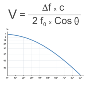
The Doppler angle of insonation
CorrectIncorrect52 of 52Question52Match the phrases on the left side with the phrases on the right side to form true statements. Click “Submit” when finished.https://usabcd.org/wp-content/uploads/FM_media/FATEadv/FATEadv_Q_M10_P21?
Sort elements
- often incurs adjustment of one or more parameters on the keyboard
- additional Doppler recordings can improve the assessment of the diastolic function
- should be used for the evaluation of the diastolic function
- is an essential part of Doppler evaluation of diastolic function in the POC context
- is essential elements for a comprehensive evaluation of diastolic dysfunction
-
When a uniform spectral Doppler curve is obtained additional optimisation
-
After evaluation of LV myocardial thickness and LA size
-
PWD but not CWD
-
E wave, A wave, E/A and deceleration time
-
Tissue Doppler and wave propagation
CorrectIncorrecterror: Content is protected !!
-