100%
Dark site-logo
Dark site-logo
  • FATE ultrasound
    • Basic FATE course
    • Advanced FATE course
    • The FATE card
  • E-learning shop
  • Workshops
  • About us
Login
  • E-learning shop
  • Workshops
  • Redeem Enrollment Code
  • Log In

LK native LD2: Advanced FATE

Instructor Image hemslars
11 Lessons
Free
This course is currently closed
Free
This course is currently closed
Course Details

LK native LD2: Advanced FATE

Instructor Image hemslars
11 Lessons
10 Quizzes
Current Status
Not Enrolled
Price
Closed
Get Started
This course is currently closed

FATE is an acronym for “Focus Assessed Transthoracic Echocardiography” and is the original focused echo protocol for all physicians. FATE is easily and quickly learned and can be applied in all clinical scenarios.

The Advanced FATE course will take you the final step in order to do a comprehensive cardiac evaluation of the critically ill patient.

Course Content

Expand All
1 Introduction to Advanced FATE
3 Topics
Expand
Lesson Content
0% Complete 0/3 Steps
FATE levels of training
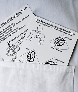
The FATE Card
Course objectives for Advanced FATE
2 Advanced FATE equipment and skills
8 Topics | 1 Quiz
Expand
Lesson Content
0% Complete 0/8 Steps
The probes used for Doppler ultrasonography
The keyboard
The keyboard - continued
Doppler gain
Doppler baseline shift
Pulse repetition frequency
Low velocity reject
Summary of Advanced FATE equipment & skills
QUIZ: Advanced FATE equipment and skills - billeder mangler
3 Inferior vena cava - IVC
16 Topics | 1 Quiz
Expand
Lesson Content
0% Complete 0/16 Steps
Position 1, the IVC view and the FATE card
Transducer placement and orientation
Transducer placement and scanning plane
Scanning plane and 2D image
Position 1: The displayed image on the screen
Position 1: IVC 2D mirror image
Position 1: IVC, anatomy of the 2D image
Obtaining the longitudinal image of the IVC from the subcostal 4 chamber view
Obtaining the longitudinal image of the IVC from the short axis view of the abdominal vessels
Measurement of the IVC diameter
IVC dimension and collapsibility correspond to the RA pressure
Using IVC dynamics for volume responsiveness
IVC diameter
Summary of IVC
QUIZ: IVC - billeder mangler nogle steder
4 Extended apical views
33 Topics | 1 Quiz
Expand
Lesson Content
0% Complete 0/33 Steps
1 - FATE position 2: Extended apical views
Position 2: Extended apical views and the FATE card
Transducer placement in apical view
Obtaining the extended apical views
FATE position 2: The apical 2 chamber view
Scanning plane and 2D Image
FATE position 2: The apical 2 chamber view
Apical 2 chamber view and the myocardial walls
Anatomy and apical 2 chamber view
Dynamic apical 2 chamber view
Obtaining the apical 2 chamber view
FATE position 2: The apical long axis view
Scanning plane and 2D image
Apical long axis view and the myocardial walls
Anatomy and apical long axis view
Dynamic apical long axis view
Obtaining the apical long axis view
FATE position 2: Display of the apical 5 chamber view
1 of 2
5 Extended parasternal views
21 Topics | 1 Quiz
Expand
Lesson Content
0% Complete 0/21 Steps
Position 3: Extended parasternal views and the FATE card
FATE position 3: Transducer placement and orientation
Extended parasternal views: scanning planes and 2D images
PSAX view mitral level: Scanning plane and 2D Image
PSAX view mitral level: Display of the scanning plane on the screen
PSAX view mitral level: Anatomy and 2D image
Dynamic parasternal short axis view at the mitral level
Obtaining the parasternal SAX view at the mitral level
PSAX view mitral level: Important points
PSAX view aorta level: Scanning plane and 2D Image
PSAX view aorta level: Display of the scanning plane on the screen
PSAX view aortic level: Pulmonary artery anatomy and 2D image
19 - PSAX view aortic level
PSAX view aortic level: Aortic valve anatomy and 2D image
Obtaining the parasternal SAX view at the aortic level
Dynamic parasternal short axis view at the aortic level
PSAX aortic level: Important points
1 of 2
6 The suprasternal view
8 Topics | 1 Quiz
Expand
Lesson Content
0% Complete 0/8 Steps
Suprasternal view
Transducer placement and orientation
Scanning plane and 2D image
Display of the scanning plane on the screen
Anatomy and 2D image
Obtaining the suprasternal view of the aortic arch
Dynamic colour flow image of the aortic arch
Summary of the suprasternal view
QUIZ: The suprasternal view
7 Basic Doppler
32 Topics | 1 Quiz
Expand
Lesson Content
0% Complete 0/32 Steps
1 - Doppler echocardiography
Doppler echocardiography
Learning objectives: Doppler echocardiography
Doppler effect
The Doppler equation - 1
The Doppler equation - 2
Angle dependency
The Doppler frequency shift
The spectral Doppler curve
Doppler modalities
Main characteristics of the Doppler modalities
Pulsed wave Doppler (PWD)
Pulsed wave Doppler (PWD) - sample volume
Continuous wave Doppler (CWD)
Continuous wave Doppler (CWD) - continuous sample line
Doppler spectral curves
Colour Doppler (CD)
Video: Colour Doppler (CD)
Video: Colour Doppler (CD) - 2
Tissue Doppler
1 of 2
8 Pressure estimation
19 Topics | 1 Quiz
Expand
Lesson Content
0% Complete 0/19 Steps
Pressure estimation with Doppler
Principle of pressure estimation with Doppler
The Bernoulli equation
The simplified Bernoulli equation
Continuous wave Doppler (CWD)
Recording the Doppler data
Analysis of the Doppler data
Pressure estimation across the aortic valve
Pitfalls: Pressure estimation across the aortic valve
Pressure estimation across the aortic valve
Correct assessment of the pressure drop across the aortic valve (5CH view)
Correct assessment of the pressure drop across the aortic valve (APLAX view)
Clinical interpretation of aortic stenosis
Pressure estimation on the right side of the heart
Pressure estimation across the tricuspid valve
Correct assessment of the pressure across a tricuspid regurgitation jet
Pressure estimation: Key points
Summary of pressure estimation
QUIZ: Pressure estimation
9 Cardiac output
12 Topics | 1 Quiz
Expand
Lesson Content
0% Complete 0/12 Steps
Principle of cardiac output estimation with Doppler ultrasound
Principle of CO estimation in a cylinder
Principle of cardiac output estimation using Doppler ultrasound
Assessment of cardiac output using Doppler ultrasound
Correct assessment of the CSA
Correct assessment of the blood flow velocity
Step-by-step assessment of cardiac output using PWD in the Left Ventricular Outflow Tract - 1
Step-by-step assessment of cardiac output using PWD in the Left Ventricular Outflow Tract - 2
Step-by-step assessment of cardiac output using PWD in the Left Ventricular Outflow Tract - 3
Cardiac output: Key points
Summary of cardiac output
QUIZ: Cardiac output in advanced FATE
10 Diastolic function
22 Topics | 1 Quiz
Expand
Lesson Content
0% Complete 0/22 Steps
General considerations for the assessment of diastolic function with Doppler echocardiography
Diastolic function
Mitral inflow with pulsed wave Doppler
Step-by-step assessment of mitral inflow blood velocities
Interpretation of the mitral diastolic inflow Doppler curve
How to measure - 1
How to measure - 2
Severity of diastolic dysfunction
Normal diastolic function
Mildly impaired diastolic function
Moderately impaired diastolic function
Severely impaired diastolic function
Tissue Doppler imaging
Tissue Doppler imaging sampling points
Step-by-step recording of tissue Doppler velocity curve in real time
Combining mitral inflow Doppler recording with TDI
How to calculate E/E'
Diastolic dysfunction in the clinical setting
LV diastolic dysfunction: The most important pitfalls
1 of 2
11 Summary of Advanced Cardiac Ultrasound (FATE)
2 Topics
Expand
Lesson Content
0% Complete 0/2 Steps
1 - Summary of the course
Join a USabcd Advanced FATE hands-on workshop
FINAL QUIZ: Advanced Cardiac Ultrasound (FATE)
  • E-learning shop
  • Workshops
  • In-hospital training
  • Medical schools
  • About us
  • Partners
  • Contact us
  • FAQ
  • facebook
  • twitter
  • linkedin
  • youtube
  • Terms of Use
  • General Conditions
  • Privacy Policy
  • Cookie Policy
USabcd ApS
Erling Jacobsens Gade 67
DK - 8240 Risskov
E-mail: [email protected]
CVR 32 87 78 85
Copyright 2017 - All Rights Reserved | Designed by WisdmLabs
error: Content is protected !!