100%
Dark site-logo
Dark site-logo
  • FATE ultrasound
    • Basic FATE course
    • Advanced FATE course
    • The FATE card
  • E-learning shop
  • Workshops
  • About us
Login
  • E-learning shop
  • Workshops
  • Redeem Enrollment Code
  • Log In

Introduction to basic ABC ultrasound for medical students

Instructor Image Peter Juhl-Olsen
9 Lessons
1507 Students
$25.00
Take this Course
$25.00
Take this Course
Course Details

Introduction to basic ABC ultrasound for medical students

Instructor Image Peter Juhl-Olsen
9 Lessons
8 Quizzes
1507 Students
Current Status
Not Enrolled
Price
€ 25
Get Started
Take this Course

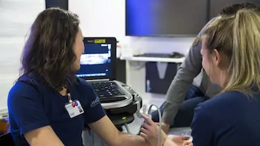
This e-course provides an introduction to the basic principles of medical ultrasound in basic lung ultrasound, focused cardiac ultrasound (FATE), the FAST protocol, ultrasound of the aorta and ultrasound for peripheral vascular access.

By completing the e-course you will be optimally prepared for the practical training

Course Content

Expand All
1 Basic Ultrasound
8 Topics | 1 Quiz
Expand
Lesson Content
0% Complete 0/8 Steps
What is a medium?
Waves
Sound wave characteristics
Attenuation
Absorption
What is ultrasound ?
Transmission and penetration
Reflection
MedStud QUIZ - Basic ultrasound
2 Equipment and Skills
13 Topics | 1 Quiz
Expand
Lesson Content
0% Complete 0/13 Steps
Preparation of the ultrasound (US) system
The ultrasound transducer
Equipment and skills
Correct orientation of the probe
Image optimisation
The orientation marker on the transducer
The ultrasound beam is equivalent to a tissue slice
Acoustic coupling - the importance of ultrasound gel
Time Gain Compensation (TGC)
Sector angle
Depth: Keep all relevant details in the image
Gain: Undergain & overgain
Summary
MedStud QUIZ - Equipment and skills
3 Focused Cardiac Ultrasound
53 Topics | 1 Quiz
Expand
Lesson Content
0% Complete 0/53 Steps
Display of the sector on the screen
Manipulating the probe
Rotation
Tilting
Sliding
Full basic FATE examination
The FATE card and abbreviations
Before you start scanning
FATE position 1: The subcostal view
Position 1 and the FATE card
Position 1: Transducer placement and orientation
Position 1: Scanning plane and 2D image
Position 1: Viewing the scanning plane on the screen
Position 1: Anatomy and 2D image
Video: Cardiac structures of beating heart
Video: Obtaining the 2D image in the subcostal position
Subcostal 4 chamber view - important points
Position 2: The apical 4 chamber view (A4CH)
Position 2 and the FATE card
Position 2: Transducer placement and orientation
1 of 3
4 Focused Cardiac Ultrasound Pathology
42 Topics
Expand
Lesson Content
0% Complete 0/42 Steps
Introduction to basic cardiac pathology
Cardiac and pleural pathology - and the FATE card
The most important pathology
Dilated, poorly functioning LV
Dilated, poorly functioning LV - typical clinical conditions
Dilated, poorly functioning LV - PSAX view
Dilated, poorly functioning LV - PLAX view
Dilated, poorly functioning LV - A4CH view
Dilated, poorly functioning LV - S4CH view
Hypertrophic LV diastolic dysfunction
Hypertrophic LV diastolic dysfunction - typical clinical conditions
Hypertrophic LV with preserved LV function - S4CH view
Hypertrophic LV with preserved LV function - PSAX view
Hypertrophic LV - PLAX view
Hypertrophic LV - AP4CH view
Pericardial effusion (cardiac tamponade) - characteristics
Cardiac tamponade
Pericardial effusion - PLAX view
Pericardial effusion - PSAX view
Pericardial effusion - A4CH view
1 of 3
5 FAST
25 Topics | 1 Quiz
Expand
Lesson Content
0% Complete 0/25 Steps
The objective of the FAST and eFAST examination
Advantages of FAST and clinical applications
Overview of FAST relevant anatomy and sonoanatomy
Free fluid and relation to the anatomical structures
The 4 FAST views
Subcostal-view: Transducer placement and scanning plane
Subcostal-view: Display of the scanning plane on the screen
Video: Obtaining the subcostal view
RUQ-view: Transducer placement and scanning plane
RUQ view: Anatomy and target view
Video: Obtaining the right upper quadrant (RUQ) view
LUQ view: Transducer placement and scanning plane
LUQ view: Anatomy and target view
Obtaining the left upper quadrant (LUQ) view
P (pelvic) views
P view - transverse: Transducer placement and scanning plane
P view - transverse: Anatomy and target view
Video: Normal examination of the transverse P view
P view - longitudinal: Transducer placement and scanning plane
P view - longitudinal: Anatomy and target view
1 of 2
6 Lung Ultrasound
31 Topics | 1 Quiz
Expand
Lesson Content
0% Complete 0/31 Steps
Normal lung and chest wall anatomy
Ultrasound presentation of the chest wall
The ”bat sign”
Transducers for lung ultrasound
Examination technique
Lung ultrasound for common diagnostics
The ”lung sliding” sign
Lung pulse
Comparing ”lung sliding” and ”lung pulse”
B-lines (Comet tails)
B-lines - appearance with two different kinds of transducers
Video: B-lines
Signs used for exclusion and diagnosis of pneumothorax (PTX)
Pleural sinus and the diaphragm - overview
Left diaphragm and pleural sinus
Right diaphragm and pleural sinus
Pleural effusion: The focused question
Normal pleura and pleural effusion on the right side
Lung ultrasound for diagnosis of interstitial syndrome
B-line artefacts
1 of 2
7 Aorta Ultrasound
6 Topics | 1 Quiz
Expand
Lesson Content
0% Complete 0/6 Steps
Anatomy of the abdominal aorta - frontal view
How to position the patient and the probe
Cross-sectional view of the abdominal aorta
Measuring the diameter of the abdominal aorta
Longitudinal US scan: the superior mesenteric artery and the celiac trunk
Abdominal aortic aneurysm
MedStud QUIZ - Aorta ultrasound
8 Ultrasound for Peripheral Vascular Access
24 Topics | 1 Quiz
Expand
Lesson Content
0% Complete 0/24 Steps
Why use ultrasound for peripheral vascular access?
How to learn US guided vascular access
Transducer for vascular access
Veins used for US-guided vascular access
Arteries used for US-guided vascular access
Blood vessel appearance on the ultrasound screen
Appearance of veins and arteries on the ultrasound screen
Compression with the transducer
Importance of diameter and depth of veins
Measuring diameter and depth below the skin surface
Appropriate length of an intravenous catheter
Introduction to ultrasound needle guidance
How does the needle tip look on the screen?
Dynamic needle tip positioning
Needle manipulation: Left/Right
Needle manipulation: Up/Down
Place needle using DNTP
Guidance markers
Patient procedures
Aseptic procedure
1 of 2
9 Course evaluation
Quiz: Introduction to ABC Ultrasound for Medical Students
  • E-learning shop
  • Workshops
  • In-hospital training
  • Medical schools
  • About us
  • Partners
  • Contact us
  • FAQ
  • facebook
  • twitter
  • linkedin
  • youtube
  • Terms of Use
  • General Conditions
  • Privacy Policy
  • Cookie Policy
USabcd ApS
Erling Jacobsens Gade 67
DK - 8240 Risskov
E-mail: [email protected]
CVR 32 87 78 85
Copyright 2017 - All Rights Reserved | Designed by WisdmLabs
error: Content is protected !!