100%
Dark site-logo
Dark site-logo
  • FATE ultrasound
    • Basic FATE course
    • Advanced FATE course
    • The FATE card
  • E-learning shop
  • Workshops
  • About us
Login
  • E-learning shop
  • Workshops
  • Redeem Enrollment Code
  • Log In

Focused Cardiac Ultrasound (FATE)

Instructor Image Erik Sloth
10 Lessons
1651 Students
$50.00
This course is currently closed
$50.00
This course is currently closed
Course Details

Focused Cardiac Ultrasound (FATE)

Instructor Image Erik Sloth
10 Lessons
9 Quizzes
1651 Students
Current Status
Not Enrolled
Price
€ 50
Get Started
This course is currently closed

Take this FATE – Basic Cardiac Ultrasound course and improve your patient care.

FATE view
FATE image
FATE cartoon

The FATE Cardiac Ultrasound Course – Focus Assessed Transthoracic Echocardiography (FATE) is the original focused cardiac ultrasound protocol practiced since 1989.

The FATE protocol is quick and easy to learn and can be applied in all clinical scenarios: the perioperative, the pre- and in-hospital, the intensive care and the emergency settings. 

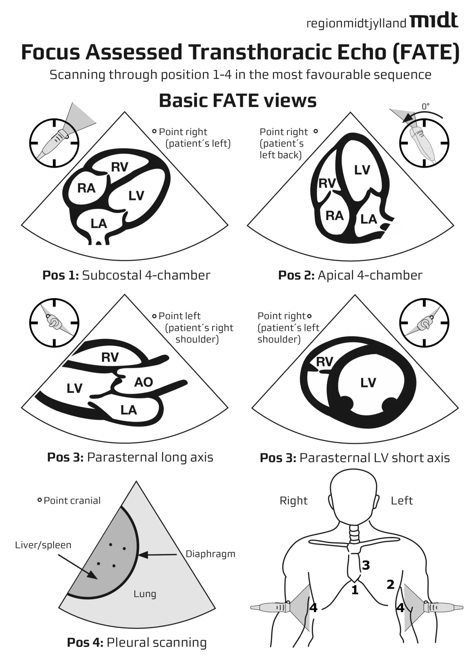
A free sample of the course – ‘Postion 1: The subcostal view’ can be viewed HERE

This is what you’ll learn:

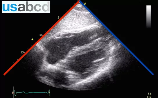
  • The normal sonographic features of heart and pleura
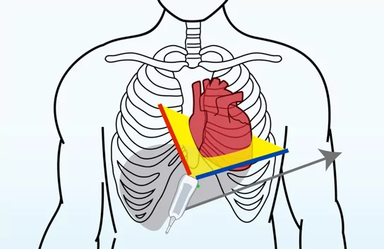
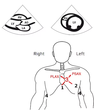
  • How to obtain the 6 basic FATE views
  • Eye-balling of right and left ventricular function
  • M-mode for assessing cardiac dimensions and function
  • The most important pathologies
  • How to interpret the echocardiographic findings

These basic echocardiographic skills are useful for physicians who are not specialists of cardiology, e.g. anaesthesiologists, intensivists, emergency physicians, internal medicine physicians, surgeons, general practitioners. In fact, all healthcare professionals dealing with hemodynamic problems in their daily clinic can benefit from knowing the FATE protocol.

Please allow 6-8 hours to complete the e-course.

Your patients will benefit from your new knowledge because FATE:

  • is quick and easy to perform
  • can be performed in the sitting position and in any location
  • is applicable in all clinical scenarios, e.g. pre- peri- and post-operatively, in intensive care, emergencies, resuscitation and so on.

Hands-on training

After completing the e-course you are ready to start getting experience in using the ultrasound machine. Completion is mandatory if you are going to attend a USabcd hands-on FATE – Basic Cardiac Ultrasound workshop. You can see all our hands-on courses in our workshop calendar.

The British Intensive Care Society has FICE approved the FATE protocol. Likewise, the FATE e-course covers the entire FOCUS curriculum. In other words, you can use the FATE – Basic Cardiac Ultrasound e-course as preparation for FICE, FOCUS and any other Point of Care cardiac ultrasound courses as it covers all learning objectives.

The FATE card

The FATE card is a pocket guide to obtaining the cardiac views. Learn more about it here.

The author

The author of the course is Prof. Erik Sloth, who invented the original focused cardiac ultrasound protocol practiced since 1989 (FATE = Focus Assessed Transthoracic Echocardiography).

Read more

  • The FATE – Basic Cardiac Ultrasound e-course is built on the foundation of this article.
  • Description on FATE-protocol on Wikipedia.
  • Focus-Assessed Transthoracic Echocardiography: Implications in Perioperative and Intensive Care by Amarja Sachin Nagre in Annals of cardiac anaesthesia 22(3); Jul-Sep 2019.
  • Focused Assessed Transthoracic Echo: Bedside Ultrasound/ Point of Care Ultrasound approach to the cardiac and pleural space examination. Dr. Liz Turner, Director of Bedside Ultrasound, UCLA.
  • Focus Assessed Transthoracic Echocardiography (FATE) Courses at HSS. Dr. Stephen Haskins, Department of Anesthesiology at Hospital for Special Surgery offers annual point-of-care ultrasound/Basic focus assessed transthoracic echocardiography (FATE) courses for anesthesiologists around the country.

FATE course in other languages

We have the FATE course in other languages too – German, Spanish, Portuguese and Japanese. Use the links below to go to those courses.

FATE course in German
FATE course in Spanish
FATE course in Portuguese
FATE course in Japanese

Course Content

Expand All
1 FATE introduction
4 Topics
Expand
Lesson Content
0% Complete 0/4 Steps
FATE levels of training
The FATE card
Course objective for Basic FATE
Your experience before this FATE course and disclaimer
2 Equipment and skills
18 Topics | 1 Quiz
Expand
Lesson Content
0% Complete 0/18 Steps
Equipment and skills
The ultrasound probe (transducer) for FATE
The orientation marker on the transducer
How to hold the probe properly
Manipulating the probe
Instruction in manipulation of the probe
Rotation
Tilting
Sliding
The sector and its orientation
Display of the sector on the screen
ECG
Image optimisation
Gain: Undergain & overgain
Depth: Keep all relevant details in the image
Sector angle
Time gain compensation (TGC)
Summary - Equipment and skills
FATE QUIZ: Equipment and skills
3 Standard projections
16 Topics | 1 Quiz
Expand
Lesson Content
0% Complete 0/16 Steps
Full Basic FATE examination
The FATE card and abbreviations
Before you start scanning
Scanning procedure
Position 1: The subcostal 4-chamber view (S4CH)
Position 1: Ultrasound image (S4CH)
Position 2: The apical 4-chamber view (A4CH)
Position 2: Ultrasound image (A4CH)
Position 3: Parasternal (P) views - two views
Position 3: Parasternal long axis view (PLAX)
Position 3: PLAX ultrasound image
Position 3: Parasternal short axis view (PSAX)
Position 3: PSAX ultrasound image
Position 4: Pleural views
Position 4: Conventions and use of pleural views
Summary - Standard projections
FATE QUIZ: Standard projections
4 Position 1
31 Topics | 1 Quiz
Expand
Lesson Content
0% Complete 0/31 Steps
FATE position 1: The subcostal view
Position 1 and the FATE card
Transducer placement and orientation
Scanning plane and 2D image
Position 1: Viewing the scanning plane on the screen
Position 1: Anatomy and 2D image
Video: Cardiac structures of the beating heart
How to obtain the 2D image in the subcostal position
Video: Obtaining the 2D image in the subcostal position
Video: Subcostal 4 chamber view from 6 healthy subjects
Video: Subcostal 4 chamber view from healthy person, obtained by different experts
Blood supply - subcostal 4 chamber view
Subcostal 4 chamber view - important points
FATE position 1: The IVC view
Position 1: The IVC view and the FATE card
Transducer placement and orientation
Transducer placement and scanning plane
Scanning plane and 2D image
Position 1: The displayed image on the screen
Position 1: IVC 2D mirror image
1 of 2
5 Position 2
16 Topics | 1 Quiz
Expand
Lesson Content
0% Complete 0/16 Steps
Position 2: The apical 4 chamber view (A4CH)
Position 2 and the FATE card
Transducer placement and orientation
Scanning plane and 2D image
Position 2: Viewing the scanning plane on the screen
Anatomy and 2D image
Video: Cardiac structures of the beating heart
How to obtain the 2D image in the apical position
Video: Obtaining the 2D image in the apical position
Video: The ictus cordis (apex beat)
Video: Apical 4 chamber view from 6 healthy subjects
Video: Apical 4 chamber view from a healthy person, obtained by 3 different experts
Blood supply - apical 4 chamber view
Apical 4 chamber view - important points
Summary - Position 2
Bonus: Be happy with the apical 4 chamber view
FATE QUIZ: Position 2
6 Position 3
28 Topics | 1 Quiz
Expand
Lesson Content
0% Complete 0/28 Steps
FATE position 3: Parasternal view
Position 3 and the FATE card
Position 3: Scanning planes and two 2D images
PLAX view: Transducer placement and orientation
PLAX view: Scanning plane and 2D image
PLAX view: Viewing the scanning plane on the screen
PLAX view: Anatomy and 2D image
PLAX video: Cardiac structures of the beating heart
How to obtain the 2D image in the parasternal long axis view
PLAX video: Obtaining the 2D parasternal long axis image in position 3
PLAX video: Parasternal long axis view from 6 healthy subjects
PLAX video: View from one healthy person, obtained by different experts
Blood supply - parasternal long axis view
PLAX view: Key points
PSAX view: Transducer placement and orientation
PSAX view: Scanning plane and 2D image
PSAX view: Viewing the scanning plane on the screen
PSAX view: Anatomy and 2D image
PSAX video: Cardiac structures of the beating heart
How to obtain the 2D image in the parasternal short axis view
1 of 2
7 Position 4
10 Topics | 1 Quiz
Expand
Lesson Content
0% Complete 0/10 Steps
FATE position 4: Pleural view
Position 4 and the FATE card
Transducer placement and orientation
Scanning planes and the 2D images
Anatomy and 2D image
How to obtain the 2D image in the pleural positions
Video: Obtaining the 2D image in the pleural position
Pleural view - important points
Summary - Position 4
Bonus - be happy with pleura scanning
FATE QUIZ: Position 4
8 Cardiac function
43 Topics | 1 Quiz
Expand
Lesson Content
0% Complete 0/43 Steps
FATE and cardiac function
FATE and ventricular function
Systolic ventricular function
Diastolic ventricular function
Left ventricular systolic function
Ejection fraction (EF) as a measure of left ventricular function
EF equation pitfalls
Eyeballing
Eyeballing hypovolaemia
Confounders of interpretation of left ventricular systolic function
M-mode ultrasound
M-mode: Presentation on the ultrasound screen
Building of the M-mode image
Understanding the M-mode image
M-mode recordings in the PLAX view
M-mode measures of LV function in the PLAX view 1
M-mode measures of LV function in the PLAX view 3
Fractional shortening (FS)
Left ventricle dimensions
LV size given by the European Society of Cardiology
1 of 3
9 Cardiac pathology
41 Topics | 1 Quiz
Expand
Lesson Content
0% Complete 0/41 Steps
Pathology
Cardiac and pleural pathology - and the FATE card
The most important pathology
Dilated, poorly functioning LV
Dilated, poorly functioning LV - typical clinical conditions
Dilated, poorly functioning LV - PSAX view
Dilated, poorly functioning LV - PLAX view
Dilated, poorly functioning LV - A4CH view
Dilated, poorly functioning LV - S4CH view
Hypertrophic LV diastolic dysfunction
Hypertrophic LV diastolic dysfunction - typical clinical conditions
Hypertrophic LV with preserved LV function - S4CH view
Hypertrophic LV with preserved LV function - PSAX view
Hypertrophic LV - PLAX view
Hypertrophic LV - A4CH view
Pericardial effusion (cardiac tamponade) - characteristics
Cardiac tamponade
Pericardial effusion - PLAX view
Pericardial effusion - PSAX view
Pericardial effusion - A4CH view
1 of 3
10 FATE course summary
6 Topics
Expand
Lesson Content
0% Complete 0/6 Steps
Summary of the Basic FATE e-learning
Self assessment for FATE course
Patients benefit from your knowledge
USabcd hands-on Basic FATE workshop
Limitations
Final remarks
FINAL QUIZ: Basic Cardiac Ultrasound (FATE)
  • E-learning shop
  • Workshops
  • In-hospital training
  • Medical schools
  • About us
  • Partners
  • Contact us
  • FAQ
  • facebook
  • twitter
  • linkedin
  • youtube
  • Terms of Use
  • General Conditions
  • Privacy Policy
  • Cookie Policy
USabcd ApS
Erling Jacobsens Gade 67
DK - 8240 Risskov
E-mail: [email protected]
CVR 32 87 78 85
Copyright 2017 - All Rights Reserved | Designed by WisdmLabs
error: Content is protected !!